This product recognizes the heavy and light chains of Guinea Pig IgG.
Applications
ICC/IF, IHC, WB, Flow Cytometry
Recommended Dilutions
IF: 1-10 µg/mL, WB: 50-100 ng/mL, IHC: 1-10 µg/mL
Clonality
Polyclonal
Isotype
IgG
Conjugate
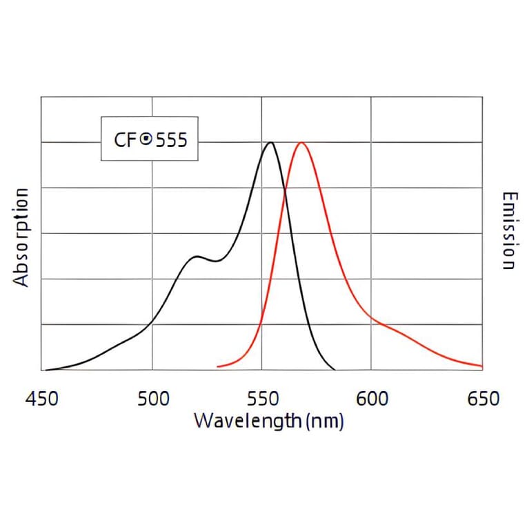
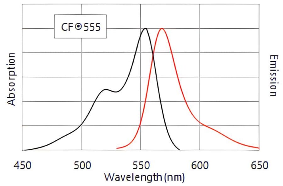
CF®555
Excitation: 555nm, Emission: 565nm
Product Form
Supplied as a liquid; 1 mg vials are lyophilized.
Concentration
2 mg/mL
Formulation
Liquid: Supplied in phosphate buffered saline (PBS) containing 50% glycerol, 2 mg/ml BSA, and 0.05% sodium azide
Storage
Shipped at +4°C. Upon delivery aliquot and store at -20°C. Avoid freeze/thaw cycles. This product is also photosensitive and should be protected from light. Should this product contain a precipitate we recommend microcentrifugation before use. CF® Dyes are guaranteed for at least 6 months from data of receipt when stored correctly.
General Notes
Looking for a specific protein conjugate to simplify your workflow? We offer a library of over 2,000 targets conjugated to your choice of CF® dye. To enquire about a custom product, contact us directly.
Disclaimer
This product is for research use only. It is not intended for diagnostic or therapeutic use.
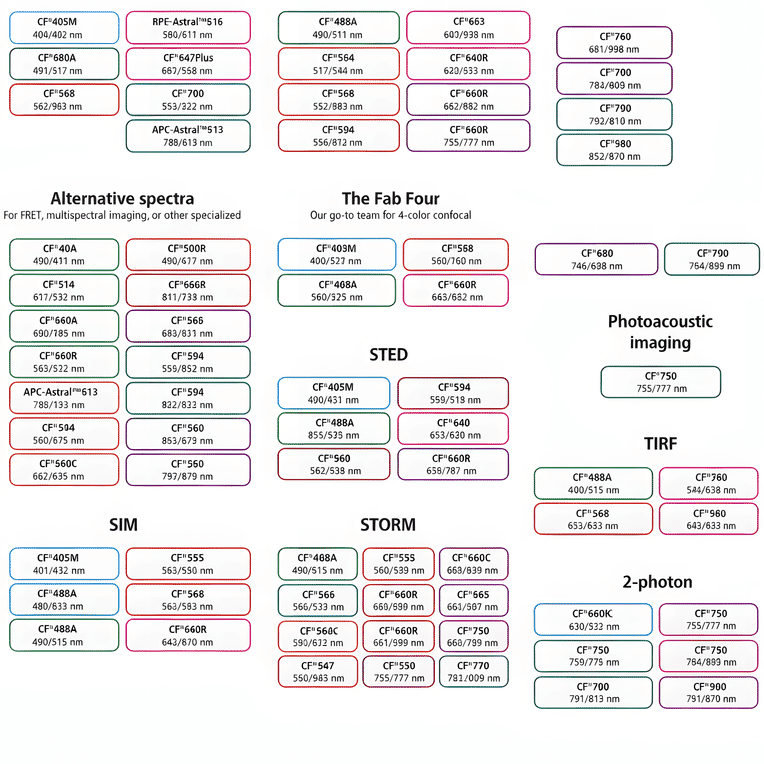
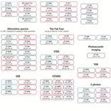
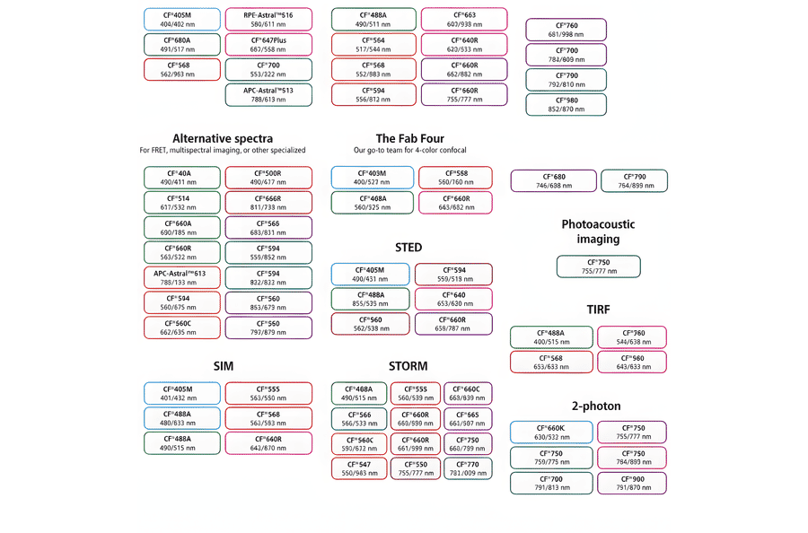
This chart summarizes commonly used CF® dyes grouped by their suitability for specific imaging modalities, including alternative spectra applications, four-color confocal imaging, near-infrared western blotting, photoacoustic imaging, STED, SIM, STORM, TIRF, and two-photon microscopy. Each dye is shown with its characteristic excitation and emission wavelengths (nm), providing a practical reference for selecting spectrally compatible dyes and optimizing multicolor experimental design across a range of fluorescence techniques.
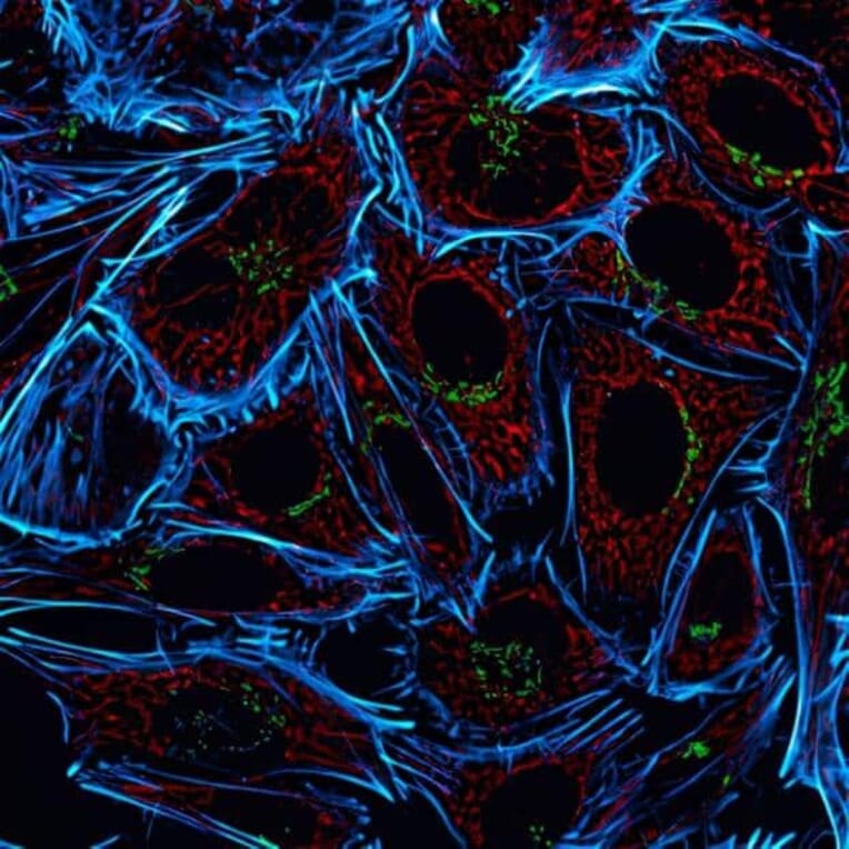
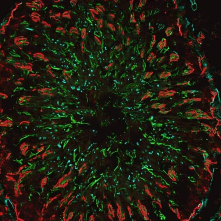
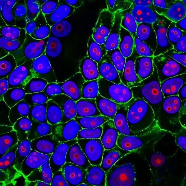
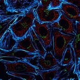
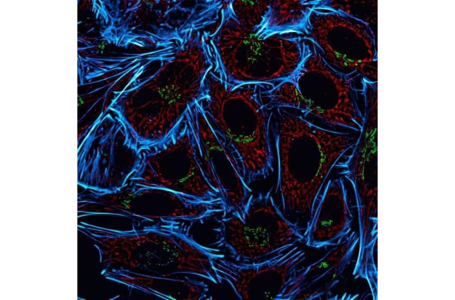
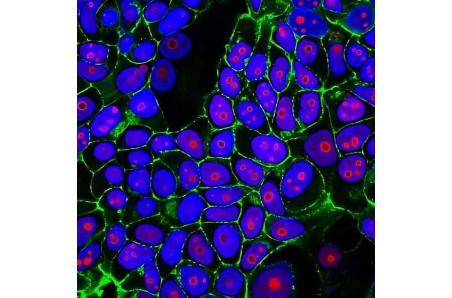
HeLa cells stained with rabbit anti-COX IV followed by CF®555 goat anti-rabbit to label mitochondria (red), mouse anti-Golgin-97 followed by CF®488A goat anti-mouse to label the Golgi complex (green), and CF®640R phalloidin to visualize actin filaments (cyan).
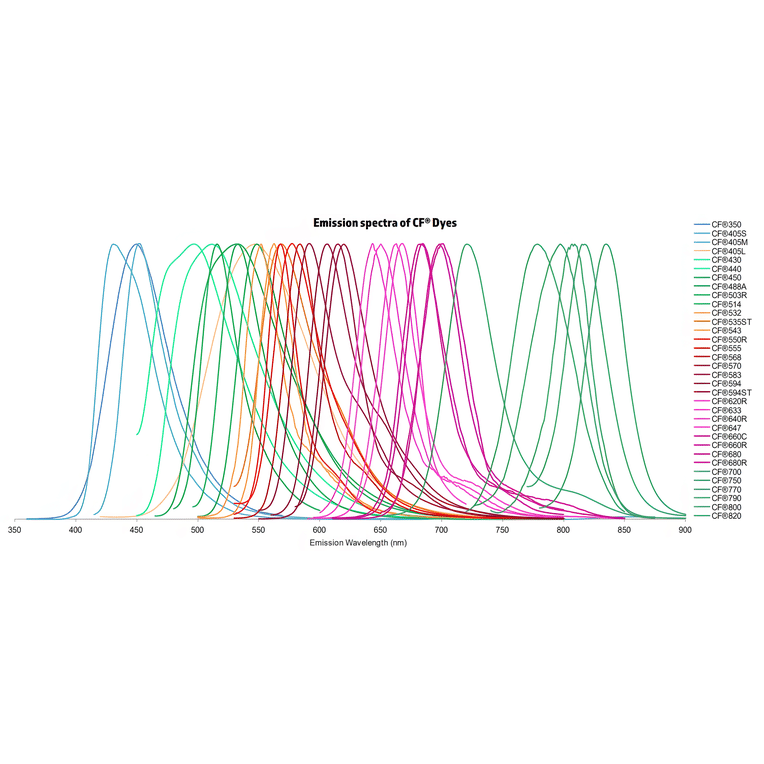
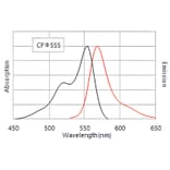
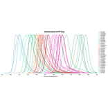
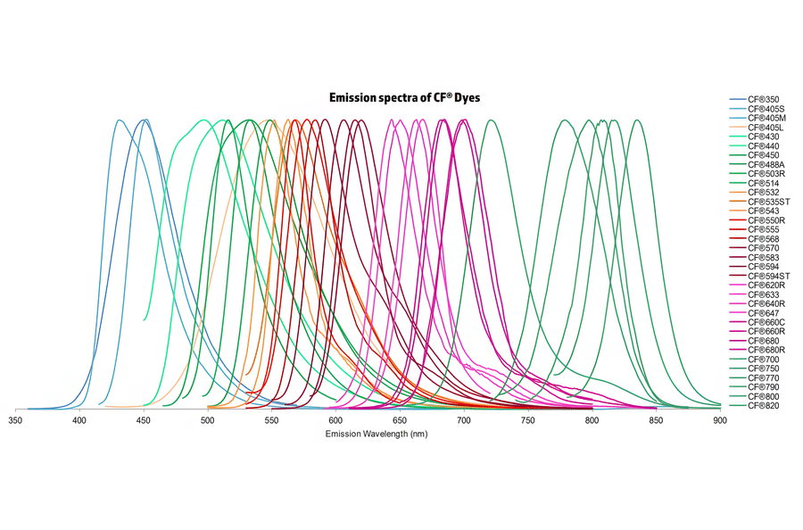
Normalized emission spectra of the CF® dye family spanning the visible to near-infrared range are shown, illustrating the spectral diversity and overlap between dyes. Curves represent relative fluorescence intensity as a function of emission wavelength (nm), with peak positions corresponding to each dye’s characteristic emission maximum. This reference highlights the broad coverage of CF® dyes for multicolor fluorescence applications and aids in selecting compatible dye combinations for imaging, flow cytometry, and other fluorescence-based assays.
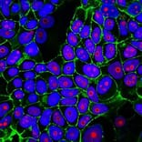
HeLa cells stained with rabbit anti-COX IV followed by CF®555 goat anti-rabbit to label mitochondria (red), mouse anti-Golgin-97 followed by CF®488A goat anti-mouse to label the Golgi complex (green), and CF®640R phalloidin to visualize actin filaments (cyan).
Publishing research using Goat Anti-Guinea Pig IgG H&L Antibody (CF®555), Cross-Adsorbed (A343943)? Please let us know so that we can list the citation on this page.