Unconjugated
TGF-β is a cytokine thought to function as a tumor promoter in advanced malignancies. In this setting, TGF-β increases cancer cell proliferation, survival, and migration, and orchestrates complex, pro-tumorigenic changes in the tumor microenvironment. Here, we find that in melanoma, integrin β1-mediated TGF-β activation may also produce tumor suppression via an altered host response. In the A375 human melanoma cell nu/nu xenograft model, we demonstrate that cell surface integrin β1-activation increases TGF-β activity, resulting in stromal activation, neo-angiogenesis and, unexpectedly for this nude mouse model, increase in the number of intra-tumoral CD8+ T lymphocytes within the tumor microenvironment. This is associated with attenuation of tumor growth and long-term survival benefit. Correspondingly, in human melanomas, TGF-β1 correlates with integrin β1/TGF-β1 activation and the expression of markers for vasculature and stromal activation. Surprisingly, this integrin β1/TGF-β1 transcriptional footprint also correlates with the expression of markers for tumor-infiltrating lymphocytes, multiple immune checkpoints and regulatory pathways, and, importantly, better long-term survival of patients. These correlations are unique to melanoma, in that we do not observe similar associations between β1 integrin/TGF-β1 activation and better long-term survival in other human tumor types. These results suggest that activation of TGF-β1 in melanoma may be associated with the generation of an anti-tumor host response that warrants further study.
In the current study, the alopecia areata gene was introduced into the C57BL/6 (B6) mouse through repeated backcrossing/intercrossing, and the allelic homozygosity of congenic AA(tj)mice (named B6.KM-AA) was verified using microsatellites. The gross appearance, growth characteristics, pathological changes in skin, and major organs of B6.KM-AA mice were observed. Counts and proportions of CD4⁺ and CD8⁺ T lymphocytes in peripheral blood were determined by flow cytometry. Results show that congenic B6.KM-AA mice were obtained after 10 generations of backcrossing/intercrossing. B6.KM-AA mice grew slower than B6 control mice and AA skin lesions were developed by four weeks of age. The number of hair follicles was reduced, but hair structures were normal. Loss of hair during disease progression was associated with CD4⁺ and CD8⁺ T lymphocytes infiltration peri-and intra-hair follicles. No pathological changes were found in other organs except for the skin. In the peripheral blood of B6.KM-AA mice, the percentage of CD4⁺ T cells was lower and percentage of CD8⁺ T cells higher than in control mice. These findings indicate that B6.KM-AA mice are characterized by a dysfunctional immune system, retarded development and T-cell infiltration mediated hair loss, making them a promising new animal model for human alopecia areata.


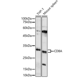
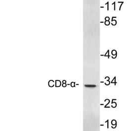
![Western Blot - Anti-CD8 alpha Antibody [ARC0329] (A306457) - Antibodies.com](https://cdn.antibodies.com/image/catalog/306/A306457_1.jpg?profile=product_alternative)
![Western Blot - Anti-CD8 alpha Antibody [ARC59319 + ARC55249] (A309931) - Antibodies.com](https://cdn.antibodies.com/image/catalog/309/A309931_1.jpg?profile=product_alternative)
![Western Blot - Anti-CD8 alpha Antibody [ARC59319] (A309962) - Antibodies.com](https://cdn.antibodies.com/image/catalog/309/A309962_1.jpg?profile=product_alternative)