Anti-Caspase 9 (A149) Anticorpo (A25826) è stato interrotto e non è più disponibile.
Visualizza tutti gli Anti-Caspase 9 Anticorpi.
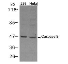
Unconjugated
Our group was the first one reporting that autophagy could be triggered by airborne fine particulate matter (PM) with a mean diameter of less than 2.5 μm (PM2.5) in human lung epithelial A549 cells, which could potentially lead to cell death. In the present study, we further explored the potential interactions between autophagy and apoptosis because it was well documented that PM2.5 could induce apoptosis in A549 cells. Much to our surprise, we found that PM2.5-exposure caused oxidative stress, resulting in activation of multiple cell death pathways in A549 cells, that is, the tumor necrosis factor-alpha (TNF-α)-induced pathway as evidenced by TNF-α secretion and activation of caspase-8 and -3, the intrinsic apoptosis pathway as evidenced by increased expression of pro-apoptotic protein Bax, decreased expression of anti-apoptotic protein Bcl-2, disruption of mitochondrial membrane potential, and activation of caspase-9 and -3, and autophagy as evidenced by an increased number of double-membrane vesicles, accompanied by increases of conversion and punctuation of microtubule-associated proteins light chain 3 (LC3) and expression of Beclin 1. It appears that reactive oxygen species (ROS) function as signaling molecules for all the three pathways because pretreatment with N-acetylcysteine, a scavenger of ROS, almost completely abolished TNF-α secretion and significantly reduced the number of apoptotic and autophagic cells. In another aspect, inhibiting autophagy with 3-methyladenine, a specific autophagy inhibitor, enhanced PM2.5-induced apoptosis and cytotoxicity. Intriguingly, neutralization of TNF-α with an anti-TNF-α special antibody not only abolished activation of caspase-8, but also drastically reduced LC3-II conversion. Thus, the present study has provided novel insights into the mechanism of cytotoxicity and even pathogenesis of diseases associated with PM2.5 exposure.
Pancreatic cancer remains the fourth most common cause of cancer-related death in the United States. Potent therapeutic strategies are urgently needed for pancreatic cancer. Cucurmosin is a novel type 1 ribosome-inactivating protein (RIP) isolated from the sarcocarp of Cucurbita moschata (pumpkin). Due to its cytotoxicity, cucurmosin can inhibit tumor cell proliferation through induction of apoptosis on tumor cells, but the specific mechanism is still unclear. We explored the function of cucurmosin in BxPC-3 pancreatic cancer cells using multiple cellular and molecular approaches such as 3-(4,5-dimethylthiazol-2-yl)-2,5-diphenyltetrazolium bromide assay, flow cytometry, reverse transcription polymerase chain reaction (RT-PCR), Western blotting and transmission electron microscopy for observing typical changes and formation of apoptotic bodies. We found that cucurmosin inhibited the proliferation of BxPC-3 cells in a time- and dose-dependent manner, and increased the cell population in the G0-G1 phase. With increasing concentration of cucurmosin, the expression of EGFR, p-PI3K, Akt, p-Akt, mTOR, p-mTOR, P70S6K-α, p-P70S6K-α, 4E-BP1 and p-4E-BP1 at the protein level was decreased, whereas the expression of p-Bad and caspase-9 was elevated. However, the mRNA expression of EGFR did not change. These findings suggest that cucurmosin can down-regulate the expression of EGFR by targeting. Cucurmosin induces the apoptosis of BxPC-3 pancreatic cancer cells via the PI3K/Akt/mTOR signaling pathway.