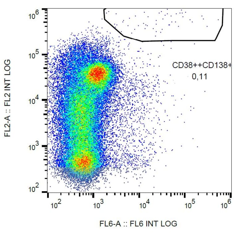
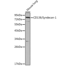
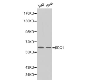
The antibody B-A38 recognizes CD138 (syndecan 1), a 65-70 kDa heparan sulfate proteoglycan expressed mainly in the epidermis and plasma cells, but also in growth factor-stimulated lymphocytes.
Applications
FC, IHC
Reactivity
Human
Immunogen
U266 human peripheral blood myeloma cell line.
Host
Mouse
Clonality
Monoclonal
Clone ID
B-A38
Isotype
IgG1
Conjugate
Unconjugated
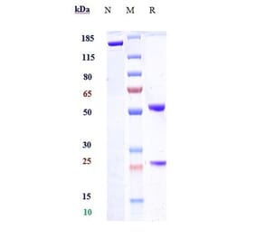
Purification
Protein A chromatography.
Concentration
1 mg/ml
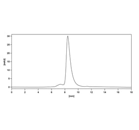
Purity
> 95% (by SDS-PAGE).
Product Form
Liquid
Formulation
Supplied in Phosphate Buffered Saline, pH 7.4, without Azide (0.2 µm filter sterilised).
Recombinant chimeric monoclonal antibody to Syndecan 1 for use as a research grade Indatuximab biosi for ELISA, Flow Cytometry, Functional Studies and in vivo Research.
Recombinant chimeric monoclonal antibody to Syndecan 1 for use as a research grade Indatuximab biosi for ELISA, Flow Cytometry, Functional Studies and in vivo Research.


![Immunofluorescence - Anti-Syndecan-1 Antibody [B-A38] (A279775) - Antibodies.com](https://cdn.antibodies.com/image/catalog/279/A279775_141706746.jpg?profile=product_alternative)
![Flow Cytometry - Anti-Syndecan-1 Antibody [DM45] - BSA and Azide free (A318650) - Antibodies.com](https://cdn.antibodies.com/image/catalog/318/A318650_1.jpg?profile=product_alternative)
![Immunohistochemistry - Anti-Syndecan 1 Antibody [SDC1/7180] (A277789) - Antibodies.com](https://cdn.antibodies.com/image/catalog/277/A277789_1.jpg?profile=product_alternative)
![Immunohistochemistry - Anti-Syndecan 1 Antibody [SDC1/7180] - BSA and Azide free (A278377) - Antibodies.com](https://cdn.antibodies.com/image/catalog/278/A278377_1.jpg?profile=product_alternative)
