Unconjugated
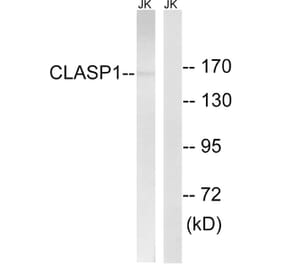
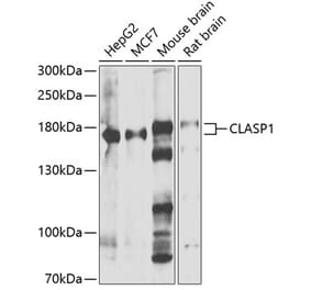
Efficient chromosome segregation during mitosis relies on the coordinated activity of molecular motors with proteins that regulate kinetochore attachments to dynamic spindle microtubules [1]. CLASPs are conserved kinetochore- and microtubule-associated proteins encoded by two paralog genes, clasp1 and clasp2, and have been previously implicated in the regulation of kinetochore microtubule dynamics [2-4]. However, it remains unknown how CLASPs work in concert with other proteins to form a functional kinetochore microtubule interface. Here we have identified mitotic interactors of human CLASP1 via a proteomic approach. Among these, the microtubule plus-end-directed motor CENP-E [5] was found to form a complex with CLASP1 that colocalizes to multiple structures of the mitotic apparatus in human cells. We found that CENP-E recruits both CLASP1 and CLASP2 to kinetochores independently of its motor activity or the presence of microtubules. Depletion of CLASPs or CENP-E by RNA interference in human cells causes a significant and comparable reduction of kinetochore microtubule poleward flux and turnover rates and rescues spindle bipolarity in Kif2a-depleted cells. We conclude that CENP-E integrates two critical functions that are important for accurate chromosome movement and spindle architecture: one relying directly on its motor activity, and the other involving the targeting of key microtubule regulators to kinetochores.
Recently, the EB1 and XMAP215/TOG families of microtubule binding proteins have been demonstrated to bind autonomously to the growing plus ends of microtubules and regulate their behaviour in in vitro systems. However, their functional redundancy or difference in cells remains obscure. Here, we compared the nanoscale distributions of EB1 and ch-TOG along microtubules using high-resolution microscopy techniques, and also their roles in microtubule organisation in interphase HeLa cells. The ch-TOG accumulation sites protruded ∼100 nm from the EB1 comets. Overexpression experiments showed that ch-TOG and EB1 did not interfere with each other's localisation, confirming that they recognise distinct regions at the ends of microtubules. While both EB1 and ch-TOG showed similar effects on microtubule plus end dynamics and additively increased microtubule dynamicity, only EB1 exhibited microtubule-cell cortex attachment activity. These observations indicate that EB1 and ch-TOG regulate microtubule organisation differently via distinct regions in the plus ends of microtubules.