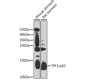
Figure 1: Western Blot - Anti-Estrogen Inducible Protein pS2 Antibody [ARC59782] (A309716)
Western blot analysis of various lysates, using Anti-Estrogen Inducible Protein pS2 Antibody [ARC59782] (A309716) at 1:1,000 dilution. The secondary antibody was Goat Anti-Rabbit IgG H&L Antibody (HRP) at 1:10,000 dilution. Lysates/proteins were present at 25µg per lane. The blocking buffer used was 3% non-fat dry milk in TBST. Detection was with a ECL Enhanced Kit (RM00021). Exposure time: 60s.
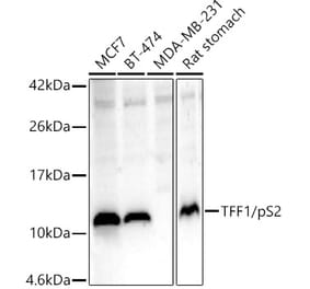
Figure 2: Western Blot - Anti-Estrogen Inducible Protein pS2 Antibody [ARC59782] (A309716)
Western blot analysis of various lysates, using Anti-Estrogen Inducible Protein pS2 Antibody [ARC59782] (A309716) at 1:1,000 dilution. The secondary antibody was Goat Anti-Rabbit IgG H&L Antibody (HRP) at 1:10,000 dilution. Lysates/proteins were present at 25µg per lane. The blocking buffer used was 3% non-fat dry milk in TBST. Detection was with a ECL Enhanced Kit (RM00021). Exposure time: 180s.
Immunofluorescence analysis of mouse spleen using Anti-Estrogen Inducible Protein pS2 Antibody [ARC59782] (A309716) at a dilution of 1:100 (40x lens). DAPI was used to stain the cell nuclei (blue).
Immunofluorescence analysis of rat stomach using Anti-Estrogen Inducible Protein pS2 Antibody [ARC59782] (A309716) at a dilution of 1:100 (40x lens). DAPI was used to stain the cell nuclei (blue).
Figure 5: Western Blot - Anti-Estrogen Inducible Protein pS2 Antibody [ARC59782] (A309716)
Immunoprecipitation analysis of 300µg extracts of MCF7 cells using 3µg of Anti-Estrogen Inducible Protein pS2 Antibody [ARC59782] (A309716). This Western blot was performed on the immunoprecipitate using Anti-Estrogen Inducible Protein pS2 Antibody [ARC59782] (A309716) at a dilution of 1:1000.


![Western Blot - Anti-Estrogen Inducible Protein pS2 Antibody [ARC59782] (A309716) - Antibodies.com](https://cdn.antibodies.com/image/catalog/309/A309716_1.jpg?profile=product_top)
![Western Blot - Anti-Estrogen Inducible Protein pS2 Antibody [ARC59782] (A309716) - Antibodies.com](https://cdn.antibodies.com/image/catalog/309/A309716_2.jpg?profile=product_top)
![Immunofluorescence - Anti-Estrogen Inducible Protein pS2 Antibody [ARC59782] (A309716) - Antibodies.com](https://cdn.antibodies.com/image/catalog/309/A309716_3.jpg?profile=product_top)
![Immunofluorescence - Anti-Estrogen Inducible Protein pS2 Antibody [ARC59782] (A309716) - Antibodies.com](https://cdn.antibodies.com/image/catalog/309/A309716_4.jpg?profile=product_top)
![Western Blot - Anti-Estrogen Inducible Protein pS2 Antibody [ARC59782] (A309716) - Antibodies.com](https://cdn.antibodies.com/image/catalog/309/A309716_5.jpg?profile=product_top)
![Western Blot - Anti-Estrogen Inducible Protein pS2 Antibody [ARC59782] (A309716) - Antibodies.com](https://cdn.antibodies.com/image/catalog/309/A309716_1.jpg?profile=product_top_thumb)
![Western Blot - Anti-Estrogen Inducible Protein pS2 Antibody [ARC59782] (A309716) - Antibodies.com](https://cdn.antibodies.com/image/catalog/309/A309716_2.jpg?profile=product_top_thumb)
![Immunofluorescence - Anti-Estrogen Inducible Protein pS2 Antibody [ARC59782] (A309716) - Antibodies.com](https://cdn.antibodies.com/image/catalog/309/A309716_3.jpg?profile=product_top_thumb)
![Immunofluorescence - Anti-Estrogen Inducible Protein pS2 Antibody [ARC59782] (A309716) - Antibodies.com](https://cdn.antibodies.com/image/catalog/309/A309716_4.jpg?profile=product_top_thumb)
![Western Blot - Anti-Estrogen Inducible Protein pS2 Antibody [ARC59782] (A309716) - Antibodies.com](https://cdn.antibodies.com/image/catalog/309/A309716_5.jpg?profile=product_top_thumb)
![Western Blot - Anti-Estrogen Inducible Protein pS2 Antibody [ARC59782] (A309716) - Antibodies.com](https://cdn.antibodies.com/image/catalog/309/A309716_1.jpg?profile=product_image)
![Western Blot - Anti-Estrogen Inducible Protein pS2 Antibody [ARC59782] (A309716) - Antibodies.com](https://cdn.antibodies.com/image/catalog/309/A309716_2.jpg?profile=product_image)
![Immunofluorescence - Anti-Estrogen Inducible Protein pS2 Antibody [ARC59782] (A309716) - Antibodies.com](https://cdn.antibodies.com/image/catalog/309/A309716_3.jpg?profile=product_image)
![Immunofluorescence - Anti-Estrogen Inducible Protein pS2 Antibody [ARC59782] (A309716) - Antibodies.com](https://cdn.antibodies.com/image/catalog/309/A309716_4.jpg?profile=product_image)
![Western Blot - Anti-Estrogen Inducible Protein pS2 Antibody [ARC59782] (A309716) - Antibodies.com](https://cdn.antibodies.com/image/catalog/309/A309716_5.jpg?profile=product_image)
![Immunohistochemistry - Anti-Estrogen Inducible Protein pS2 Antibody [TFF1/2133] (A250088) - Antibodies.com](https://cdn.antibodies.com/image/catalog/250/A250088_1.jpg?profile=product_alternative)
![Immunohistochemistry - Anti-Estrogen Inducible Protein pS2 Antibody [TFF1/2133] - BSA and Azide free (A253268) - Antibodies.com](https://cdn.antibodies.com/image/catalog/253/A253268_1.jpg?profile=product_alternative)
![Western Blot - Anti-Estrogen Inducible Protein pS2 Antibody [ARC1427] (A307065) - Antibodies.com](https://cdn.antibodies.com/image/catalog/307/A307065_1.jpg?profile=product_alternative)