Unconjugated
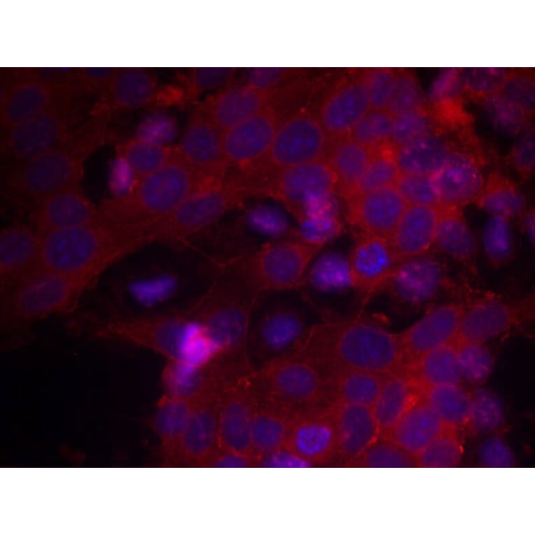
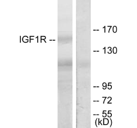
The insulin and insulin-like growth factor-1 (IGF-1) are considered to play important roles in brain development; and their cognate receptors -InsR and IGF-1R- localized within distinct brain regions including cerebellum. Using Real-Time PCR and western blot analysis, we compared the expression of InsR and IGF-1R in male and female developing rat cerebellum at P0, P7, and P14. At all time points studied, the cerebellar expression of IGF-1R, both at mRNA and protein levels was higher than that of InsR. The lowest InsR and IGF-1R mRNA and protein levels were measured in the neonate cerebellum, independent of gender. In males, the highest InsR and IGF-1R mRNA and protein expression were found at P7. InsR and IGF-1R expression increased significantly between P0 and P7, followed by a marked downregulation at P14. In contrast, in females, mRNA and protein levels of InsR and IGF-1R remain unchanged between P0 and P7, and are upregulated at P14. Therefore, peaked InsR and IGF-1R expression in female cerebelli occurred at P14. Interestingly, changes in mRNA expression and in protein levels followed the same developmental pattern, indicating that InsR and IGF-1R transcription is not subject to modulatory effects during the first 2 weeks of development. These findings indicate that there are prominent sexual differences in InsR and IGF-1R expression in the developing rat cerebellum, suggesting a probable mechanism for the control of gender differences in development and function of the cerebellum.
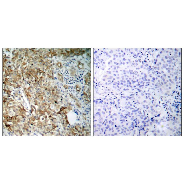
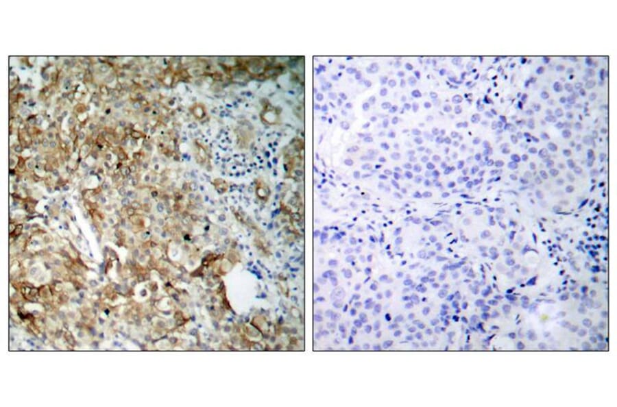
Insulin-like growth factor-1 receptor (IGF-1R) and matrix metalloproteinase-7 (MMP-7) have been reported to be related to tumor invasion and metastasis in various malignancies. The aim of this study was to evaluate the expression levels of IGF-1R and MMP-7 in resected non-small cell lung cancer (NSCLC) and to examine the relationship of such levels to clinical characteristics and survival. Expression was measured immunohistochemically. The percentage of stained cells was multiplied by the staining intensity. The sample was classified as high when the score was equal or higher than the median value or was otherwise considered to be low. High IGF-1R expression was associated with nodal metastasis and recurrence (P=0.034 and 0.006, respectively). High IGF-1R expression was associated with significantly poorer overall survival than low IGF-1R expression (P=0.011). MMP-7 expression did not significantly correlate with any clinicopathological factor. There was a trend toward slightly, but not significantly poorer survival in patients with MMP-7-high tumors than in those with MMP-7-low tumors (P=0.220). There was no significant correlation between IGF-1R expression and MMP-7 expression (P=0.184). Upon multivariate analysis, IGF-1R expression was independently related to the outcomes of patients with NSCLC. Overexpression of IGF-1R may be a useful predictor of lymph node metastasis, recurrence and post-surgical outcomes in patients with NSCLC.

![Immunohistochemistry - Anti-IGF1 Receptor Antibody [IGF1R/4667] (A277659) - Antibodies.com](https://cdn.antibodies.com/image/catalog/277/A277659_1.jpg?profile=product_alternative)
![Immunohistochemistry - Anti-IGF1 Receptor Antibody [IGF1R/4667] - BSA and Azide free (A278247) - Antibodies.com](https://cdn.antibodies.com/image/catalog/278/A278247_1.jpg?profile=product_alternative)