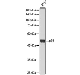
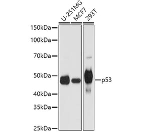
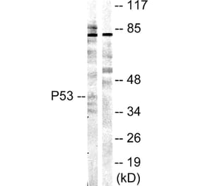
Figure 1: Western Blot - Anti-p53 Antibody [ARC50968] (A307216)
Western blot analysis of extracts from wild type(WT) and p53 knockout (KO) 293T(KO) cells, using Anti-p53 Antibody [ARC50968] (A307216) at 1:1,000 dilution. The secondary antibody was Goat Anti-Rabbit IgG H&L Antibody (HRP) at 1:10,000 dilution. Lysates/proteins were present at 25µg per lane. The blocking buffer used was 3% non-fat dry milk in TBST. Detection was with a ECL Basic Kit. Exposure time: 60s.
Immunohistochemistry analysis of paraffin-embedded human colon tissue (negative control sample) using Anti-p53 Antibody [ARC50968] (A307216) at a dilution of 1:50 (40x lens). Perform high pressure antigen retrieval with 10 mM citrate buffer pH 6.0 before commencing with IHC staining protocol.
Immunohistochemistry analysis of paraffin-embedded human colon carcinoma tissue using Anti-p53 Antibody [ARC50968] (A307216) at a dilution of 1:50 (40x lens). Perform high pressure antigen retrieval with 10 mM citrate buffer pH 6.0 before commencing with IHC staining protocol.
Immunohistochemistry analysis of paraffin-embedded human hepatocholangiocarcinoma using Anti-p53 Antibody [ARC50968] (A307216) at a dilution of 1:50 (40x lens). Perform high pressure antigen retrieval with 10 mM citrate buffer pH 6.0 before commencing with IHC staining protocol.
Immunohistochemistry analysis of paraffin-embedded human liver (negative control sample) using Anti-p53 Antibody [ARC50968] (A307216) at a dilution of 1:50 (40x lens). Perform high pressure antigen retrieval with 10 mM citrate buffer pH 6.0 before commencing with IHC staining protocol.


![Western Blot - Anti-p53 Antibody [ARC50968] (A307216) - Antibodies.com](https://cdn.antibodies.com/image/catalog/307/A307216_1.jpg?profile=product_top)
![Immunohistochemistry - Anti-p53 Antibody [ARC50968] (A307216) - Antibodies.com](https://cdn.antibodies.com/image/catalog/307/A307216_2.jpg?profile=product_top)
![Immunohistochemistry - Anti-p53 Antibody [ARC50968] (A307216) - Antibodies.com](https://cdn.antibodies.com/image/catalog/307/A307216_3.jpg?profile=product_top)
![Immunohistochemistry - Anti-p53 Antibody [ARC50968] (A307216) - Antibodies.com](https://cdn.antibodies.com/image/catalog/307/A307216_4.jpg?profile=product_top)
![Immunohistochemistry - Anti-p53 Antibody [ARC50968] (A307216) - Antibodies.com](https://cdn.antibodies.com/image/catalog/307/A307216_5.jpg?profile=product_top)
![Western Blot - Anti-p53 Antibody [ARC50968] (A307216) - Antibodies.com](https://cdn.antibodies.com/image/catalog/307/A307216_1.jpg?profile=product_top_thumb)
![Immunohistochemistry - Anti-p53 Antibody [ARC50968] (A307216) - Antibodies.com](https://cdn.antibodies.com/image/catalog/307/A307216_2.jpg?profile=product_top_thumb)
![Immunohistochemistry - Anti-p53 Antibody [ARC50968] (A307216) - Antibodies.com](https://cdn.antibodies.com/image/catalog/307/A307216_3.jpg?profile=product_top_thumb)
![Immunohistochemistry - Anti-p53 Antibody [ARC50968] (A307216) - Antibodies.com](https://cdn.antibodies.com/image/catalog/307/A307216_4.jpg?profile=product_top_thumb)
![Immunohistochemistry - Anti-p53 Antibody [ARC50968] (A307216) - Antibodies.com](https://cdn.antibodies.com/image/catalog/307/A307216_5.jpg?profile=product_top_thumb)
![Western Blot - Anti-p53 Antibody [ARC50968] (A307216) - Antibodies.com](https://cdn.antibodies.com/image/catalog/307/A307216_1.jpg?profile=product_image)
![Immunohistochemistry - Anti-p53 Antibody [ARC50968] (A307216) - Antibodies.com](https://cdn.antibodies.com/image/catalog/307/A307216_2.jpg?profile=product_image)
![Immunohistochemistry - Anti-p53 Antibody [ARC50968] (A307216) - Antibodies.com](https://cdn.antibodies.com/image/catalog/307/A307216_3.jpg?profile=product_image)
![Immunohistochemistry - Anti-p53 Antibody [ARC50968] (A307216) - Antibodies.com](https://cdn.antibodies.com/image/catalog/307/A307216_4.jpg?profile=product_image)
![Immunohistochemistry - Anti-p53 Antibody [ARC50968] (A307216) - Antibodies.com](https://cdn.antibodies.com/image/catalog/307/A307216_5.jpg?profile=product_image)
![Immunohistochemistry - Anti-p53 Antibody [rBP53-12] (A250176) - Antibodies.com](https://cdn.antibodies.com/image/catalog/250/A250176_1.jpg?profile=product_alternative)
![Immunohistochemistry - Anti-p53 Antibody [TP53/2092R] (A250192) - Antibodies.com](https://cdn.antibodies.com/image/catalog/250/A250192_1.jpg?profile=product_alternative)