Supplied in Phosphate Buffered Saline, pH 7.3, with 50% Glycerol and 0.01% Thiomersal.
Conservazione
Shipped at 4°C. Upon delivery aliquot and store at -20°C. Avoid freeze / thaw cycles.
Sinonimi
48 kDa protein, Arrestin, Arrestin 1, ARRS_HUMAN, Retinal S antigen (48 KDa protein), Retinal S-antigen, Rod photoreceptor arrestin, RP47, S antigen, S antigen retina and pineal gland (arrestin), S arrestin, S-AG, S-arrestin, SAG
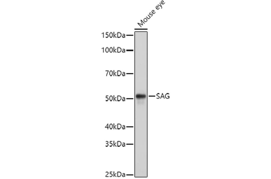
Figure 1: Western Blot - Anti-Retinal S antigen Antibody (A90119)
Western blot analysis of extracts of Mouse eye cells, using Anti-Retinal S antigen Antibody (A90119) at 1:1,000 dilution. The secondary antibody was Goat Anti-Rabbit IgG H&L Antibody (HRP) at 1:10,000 dilution. Lysates/proteins were present at 25µg per lane. The blocking buffer used was 3% non-fat dry milk in TBST. Detection was with a ECL Basic Kit. Exposure time: 1s.
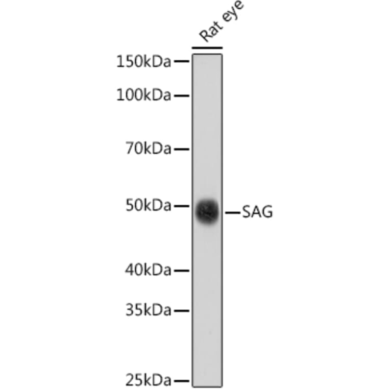
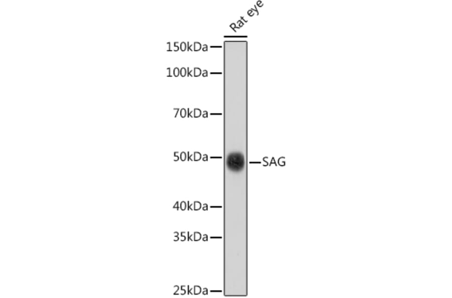
Figure 2: Western Blot - Anti-Retinal S antigen Antibody (A90119)
Western blot analysis of extracts of Rat eye cells, using Anti-Retinal S antigen Antibody (A90119) at 1:1,000 dilution. The secondary antibody was Goat Anti-Rabbit IgG H&L Antibody (HRP) at 1:10,000 dilution. Lysates/proteins were present at 25µg per lane. The blocking buffer used was 3% non-fat dry milk in TBST. Detection was with a ECL Basic Kit. Exposure time: 30s.
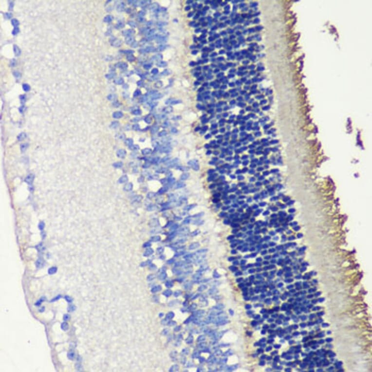
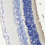
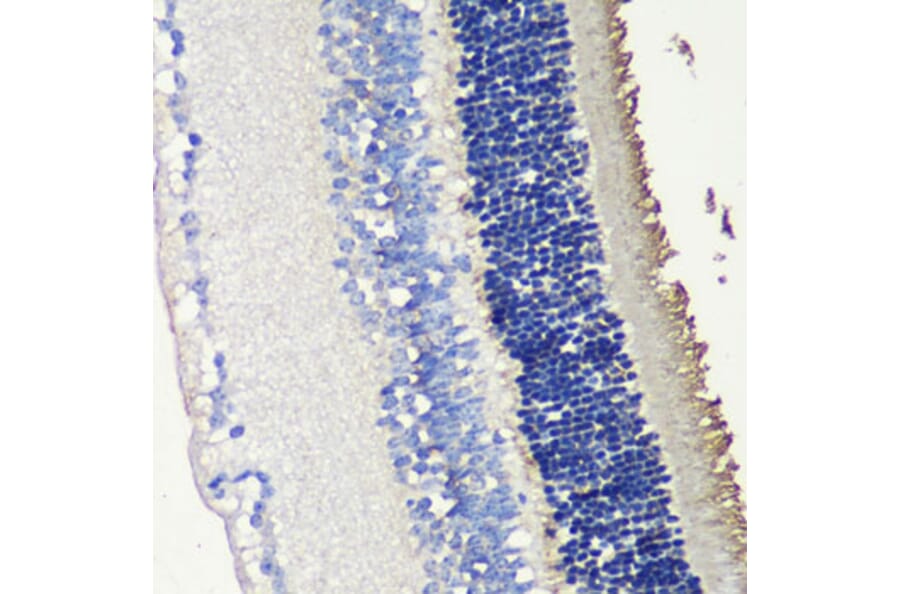
Figure 3: Immunohistochemistry - Anti-Retinal S antigen Antibody (A90119)
Immunohistochemistry analysis of paraffin-embedded mouse retina using Anti-Retinal S antigen Antibody (A90119) at a dilution of 1:200 (40x lens). Perform microwave antigen retrieval with 10 mM PBS buffer pH 7.2 before commencing with IHC staining protocol.
Prodotti alternativi a Anti-Retinal S antigen Anticorpo (A90119)