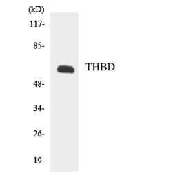
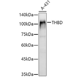
Figure 1: Western Blot - Anti-Thrombomodulin Antibody [ARC52599 + ARC52600] (A309567)
Western blot analysis of extracts of A-431, using Anti-Thrombomodulin Antibody [ARC52599 + ARC52600] (A309567) at 1:5,000 dilution. The secondary antibody was Goat Anti-Rabbit IgG H&L Antibody (HRP) at 1:10,000 dilution. Lysates/proteins were present at 25µg per lane. The blocking buffer used was 3% non-fat dry milk in TBST. Detection was with a ECL Basic Kit. Exposure time: 1s.
Immunohistochemistry analysis of paraffin-embedded human lung squamous carcinoma tissue using Anti-Thrombomodulin Antibody [ARC52599 + ARC52600] (A309567) at a dilution of 1:500 (40x lens). Perform high pressure antigen retrieval with 10 mM citrate buffer pH 6.0 before commencing with IHC staining protocol.
Immunohistochemistry analysis of paraffin-embedded human lung using Anti-Thrombomodulin Antibody [ARC52599 + ARC52600] (A309567) at a dilution of 1:500 (40x lens). Perform high pressure antigen retrieval with 10 mM citrate buffer pH 6.0 before commencing with IHC staining protocol.
Immunohistochemistry analysis of paraffin-embedded human tonsil using Anti-Thrombomodulin Antibody [ARC52599 + ARC52600] (A309567) at a dilution of 1:500 (40x lens). Perform high pressure antigen retrieval with 10 mM citrate buffer pH 6.0 before commencing with IHC staining protocol.
Immunofluorescence analysis of A431 using Anti-Thrombomodulin Antibody [ARC52599 + ARC52600] (A309567) at a dilution of 1:100 (40x lens). DAPI was used to stain the cell nuclei (blue).
Immunofluorescence analysis of THP-1 using Anti-Thrombomodulin Antibody [ARC52599 + ARC52600] (A309567) at a dilution of 1:100 (40x lens). DAPI was used to stain the cell nuclei (blue).


![Western Blot - Anti-Thrombomodulin Antibody [ARC52599 + ARC52600] (A309567) - Antibodies.com](https://cdn.antibodies.com/image/catalog/309/A309567_1.jpg?profile=product_top)
![Immunohistochemistry - Anti-Thrombomodulin Antibody [ARC52599 + ARC52600] (A309567) - Antibodies.com](https://cdn.antibodies.com/image/catalog/309/A309567_2.jpg?profile=product_top)
![Immunohistochemistry - Anti-Thrombomodulin Antibody [ARC52599 + ARC52600] (A309567) - Antibodies.com](https://cdn.antibodies.com/image/catalog/309/A309567_3.jpg?profile=product_top)
![Immunohistochemistry - Anti-Thrombomodulin Antibody [ARC52599 + ARC52600] (A309567) - Antibodies.com](https://cdn.antibodies.com/image/catalog/309/A309567_4.jpg?profile=product_top)
![Immunofluorescence - Anti-Thrombomodulin Antibody [ARC52599 + ARC52600] (A309567) - Antibodies.com](https://cdn.antibodies.com/image/catalog/309/A309567_5.jpg?profile=product_top)
![Immunofluorescence - Anti-Thrombomodulin Antibody [ARC52599 + ARC52600] (A309567) - Antibodies.com](https://cdn.antibodies.com/image/catalog/309/A309567_6.jpg?profile=product_top)
![Western Blot - Anti-Thrombomodulin Antibody [ARC52599 + ARC52600] (A309567) - Antibodies.com](https://cdn.antibodies.com/image/catalog/309/A309567_1.jpg?profile=product_top_thumb)
![Immunohistochemistry - Anti-Thrombomodulin Antibody [ARC52599 + ARC52600] (A309567) - Antibodies.com](https://cdn.antibodies.com/image/catalog/309/A309567_2.jpg?profile=product_top_thumb)
![Immunohistochemistry - Anti-Thrombomodulin Antibody [ARC52599 + ARC52600] (A309567) - Antibodies.com](https://cdn.antibodies.com/image/catalog/309/A309567_3.jpg?profile=product_top_thumb)
![Immunohistochemistry - Anti-Thrombomodulin Antibody [ARC52599 + ARC52600] (A309567) - Antibodies.com](https://cdn.antibodies.com/image/catalog/309/A309567_4.jpg?profile=product_top_thumb)
![Immunofluorescence - Anti-Thrombomodulin Antibody [ARC52599 + ARC52600] (A309567) - Antibodies.com](https://cdn.antibodies.com/image/catalog/309/A309567_5.jpg?profile=product_top_thumb)
![Immunofluorescence - Anti-Thrombomodulin Antibody [ARC52599 + ARC52600] (A309567) - Antibodies.com](https://cdn.antibodies.com/image/catalog/309/A309567_6.jpg?profile=product_top_thumb)
![Western Blot - Anti-Thrombomodulin Antibody [ARC52599 + ARC52600] (A309567) - Antibodies.com](https://cdn.antibodies.com/image/catalog/309/A309567_1.jpg?profile=product_image)
![Immunohistochemistry - Anti-Thrombomodulin Antibody [ARC52599 + ARC52600] (A309567) - Antibodies.com](https://cdn.antibodies.com/image/catalog/309/A309567_2.jpg?profile=product_image)
![Immunohistochemistry - Anti-Thrombomodulin Antibody [ARC52599 + ARC52600] (A309567) - Antibodies.com](https://cdn.antibodies.com/image/catalog/309/A309567_3.jpg?profile=product_image)
![Immunohistochemistry - Anti-Thrombomodulin Antibody [ARC52599 + ARC52600] (A309567) - Antibodies.com](https://cdn.antibodies.com/image/catalog/309/A309567_4.jpg?profile=product_image)
![Immunofluorescence - Anti-Thrombomodulin Antibody [ARC52599 + ARC52600] (A309567) - Antibodies.com](https://cdn.antibodies.com/image/catalog/309/A309567_5.jpg?profile=product_image)
![Immunofluorescence - Anti-Thrombomodulin Antibody [ARC52599 + ARC52600] (A309567) - Antibodies.com](https://cdn.antibodies.com/image/catalog/309/A309567_6.jpg?profile=product_image)
![Immunohistochemistry - Anti-Thrombomodulin Antibody [THBD/1591] (A250124) - Antibodies.com](https://cdn.antibodies.com/image/catalog/250/A250124_1.jpg?profile=product_alternative)
![Immunohistochemistry - Anti-Thrombomodulin Antibody [THBD/1591] - BSA and Azide free (A253304) - Antibodies.com](https://cdn.antibodies.com/image/catalog/253/A253304_1.jpg?profile=product_alternative)
![Immunohistochemistry - Anti-Thrombomodulin Antibody [rTHBD/1591] (A250126) - Antibodies.com](https://cdn.antibodies.com/image/catalog/250/A250126_1.jpg?profile=product_alternative)
![Immunohistochemistry - Anti-Thrombomodulin Antibody [THBD/1782] - BSA and Azide free (A253305) - Antibodies.com](https://cdn.antibodies.com/image/catalog/253/A253305_1.jpg?profile=product_alternative)
![Immunohistochemistry - Anti-Thrombomodulin Antibody [rTHBD/1591] - BSA and Azide free (A253306) - Antibodies.com](https://cdn.antibodies.com/image/catalog/253/A253306_1.jpg?profile=product_alternative)
![Immunohistochemistry - Anti-Thrombomodulin Antibody [THBD/1782] (A250125) - Antibodies.com](https://cdn.antibodies.com/image/catalog/250/A250125_1.jpg?profile=product_alternative)