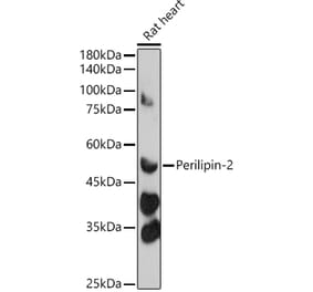
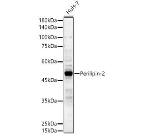
Figure 1: Western Blot - Anti-ADFP Antibody [ARC51735] (A305635)
Western blot analysis of extracts of various cell lines, using Anti-ADFP Antibody [ARC51735] (A305635) at 1:1,000 dilution. The secondary antibody was Goat Anti-Rabbit IgG H&L Antibody (HRP) at 1:10,000 dilution. Lysates/proteins were present at 25µg per lane. The blocking buffer used was 3% non-fat dry milk in TBST. Detection was with a ECL Basic Kit. Exposure time: 10s.
Immunohistochemistry analysis of paraffin-embedded human colon carcinoma tissue using Anti-ADFP Antibody [ARC51735] (A305635) at a dilution of 1:1,000 (40x lens). Perform high pressure antigen retrieval with 10 mM citrate buffer pH 6.0 before commencing with IHC staining protocol.
Immunohistochemistry analysis of paraffin-embedded human liver using Anti-ADFP Antibody [ARC51735] (A305635) at a dilution of 1:1,000 (40x lens). Perform high pressure antigen retrieval with 10 mM citrate buffer pH 6.0 before commencing with IHC staining protocol.
Immunofluorescence analysis of mouse brown adipose using Anti-ADFP Antibody [ARC51735] (A305635) at a dilution of 1:100(40x lens). DAPI was used to stain the cell nuclei (blue).
Immunofluorescence analysis of HepG2 cells using Anti-ADFP Antibody [ARC51735] (A305635) at a dilution of 1:100(40x lens). DAPI was used to stain the cell nuclei (blue).


![Western Blot - Anti-ADFP Antibody [ARC51735] (A305635) - Antibodies.com](https://cdn.antibodies.com/image/catalog/305/A305635_1.jpg?profile=product_top)
![Immunohistochemistry - Anti-ADFP Antibody [ARC51735] (A305635) - Antibodies.com](https://cdn.antibodies.com/image/catalog/305/A305635_2.jpg?profile=product_top)
![Immunohistochemistry - Anti-ADFP Antibody [ARC51735] (A305635) - Antibodies.com](https://cdn.antibodies.com/image/catalog/305/A305635_3.jpg?profile=product_top)
![Immunofluorescence - Anti-ADFP Antibody [ARC51735] (A305635) - Antibodies.com](https://cdn.antibodies.com/image/catalog/305/A305635_4.jpg?profile=product_top)
![Immunofluorescence - Anti-ADFP Antibody [ARC51735] (A305635) - Antibodies.com](https://cdn.antibodies.com/image/catalog/305/A305635_5.jpg?profile=product_top)
![Western Blot - Anti-ADFP Antibody [ARC51735] (A305635) - Antibodies.com](https://cdn.antibodies.com/image/catalog/305/A305635_1.jpg?profile=product_top_thumb)
![Immunohistochemistry - Anti-ADFP Antibody [ARC51735] (A305635) - Antibodies.com](https://cdn.antibodies.com/image/catalog/305/A305635_2.jpg?profile=product_top_thumb)
![Immunohistochemistry - Anti-ADFP Antibody [ARC51735] (A305635) - Antibodies.com](https://cdn.antibodies.com/image/catalog/305/A305635_3.jpg?profile=product_top_thumb)
![Immunofluorescence - Anti-ADFP Antibody [ARC51735] (A305635) - Antibodies.com](https://cdn.antibodies.com/image/catalog/305/A305635_4.jpg?profile=product_top_thumb)
![Immunofluorescence - Anti-ADFP Antibody [ARC51735] (A305635) - Antibodies.com](https://cdn.antibodies.com/image/catalog/305/A305635_5.jpg?profile=product_top_thumb)
![Western Blot - Anti-ADFP Antibody [ARC51735] (A305635) - Antibodies.com](https://cdn.antibodies.com/image/catalog/305/A305635_1.jpg?profile=product_image)
![Immunohistochemistry - Anti-ADFP Antibody [ARC51735] (A305635) - Antibodies.com](https://cdn.antibodies.com/image/catalog/305/A305635_2.jpg?profile=product_image)
![Immunohistochemistry - Anti-ADFP Antibody [ARC51735] (A305635) - Antibodies.com](https://cdn.antibodies.com/image/catalog/305/A305635_3.jpg?profile=product_image)
![Immunofluorescence - Anti-ADFP Antibody [ARC51735] (A305635) - Antibodies.com](https://cdn.antibodies.com/image/catalog/305/A305635_4.jpg?profile=product_image)
![Immunofluorescence - Anti-ADFP Antibody [ARC51735] (A305635) - Antibodies.com](https://cdn.antibodies.com/image/catalog/305/A305635_5.jpg?profile=product_image)
![Western Blot - Anti-ADFP Antibody [ARC51733] (A305636) - Antibodies.com](https://cdn.antibodies.com/image/catalog/305/A305636_1.jpg?profile=product_alternative)
![Immunohistochemistry - Anti-Adipophilin Antibody [ADFP/2755R] - BSA and Azide free (A251418) - Antibodies.com](https://cdn.antibodies.com/image/catalog/251/A251419_1.jpg?profile=product_alternative)
![Western Blot - Anti-Adipophilin Antibody [ADFP/1365] (A248231) - Antibodies.com](https://cdn.antibodies.com/image/catalog/248/A248231_1.jpg?profile=product_alternative)
![Western Blot - Anti-Adipophilin Antibody [ADFP/1365] - BSA and Azide free (A251414) - Antibodies.com](https://cdn.antibodies.com/image/catalog/251/A251414_1.jpg?profile=product_alternative)
![Immunohistochemistry - Anti-Adipophilin Antibody [ADFP/2755R] (A248235) - Antibodies.com](https://cdn.antibodies.com/image/catalog/248/A248236_1.jpg?profile=product_alternative)
![Immunohistochemistry - Anti-Adipophilin Antibody [ADFP/1366] (A248232) - Antibodies.com](https://cdn.antibodies.com/image/catalog/248/A248232_1.jpg?profile=product_alternative)
![Immunohistochemistry - Anti-Adipophilin Antibody [ADFP/1366] - BSA and Azide free (A251415) - Antibodies.com](https://cdn.antibodies.com/image/catalog/251/A251415_1.jpg?profile=product_alternative)
![Immunohistochemistry - Anti-Adipophilin Antibody [rADFP/1493] - BSA and Azide free (A251417) - Antibodies.com](https://cdn.antibodies.com/image/catalog/251/A251418_1.jpg?profile=product_alternative)
![Immunohistochemistry - Anti-Adipophilin Antibody [ADFP/1493] (A248232) - Antibodies.com](https://cdn.antibodies.com/image/catalog/248/A248233_1.jpg?profile=product_alternative)
![Immunohistochemistry - Anti-Adipophilin Antibody [ADFP/1493] - BSA and Azide free (A251415) - Antibodies.com](https://cdn.antibodies.com/image/catalog/251/A251416_1.jpg?profile=product_alternative)