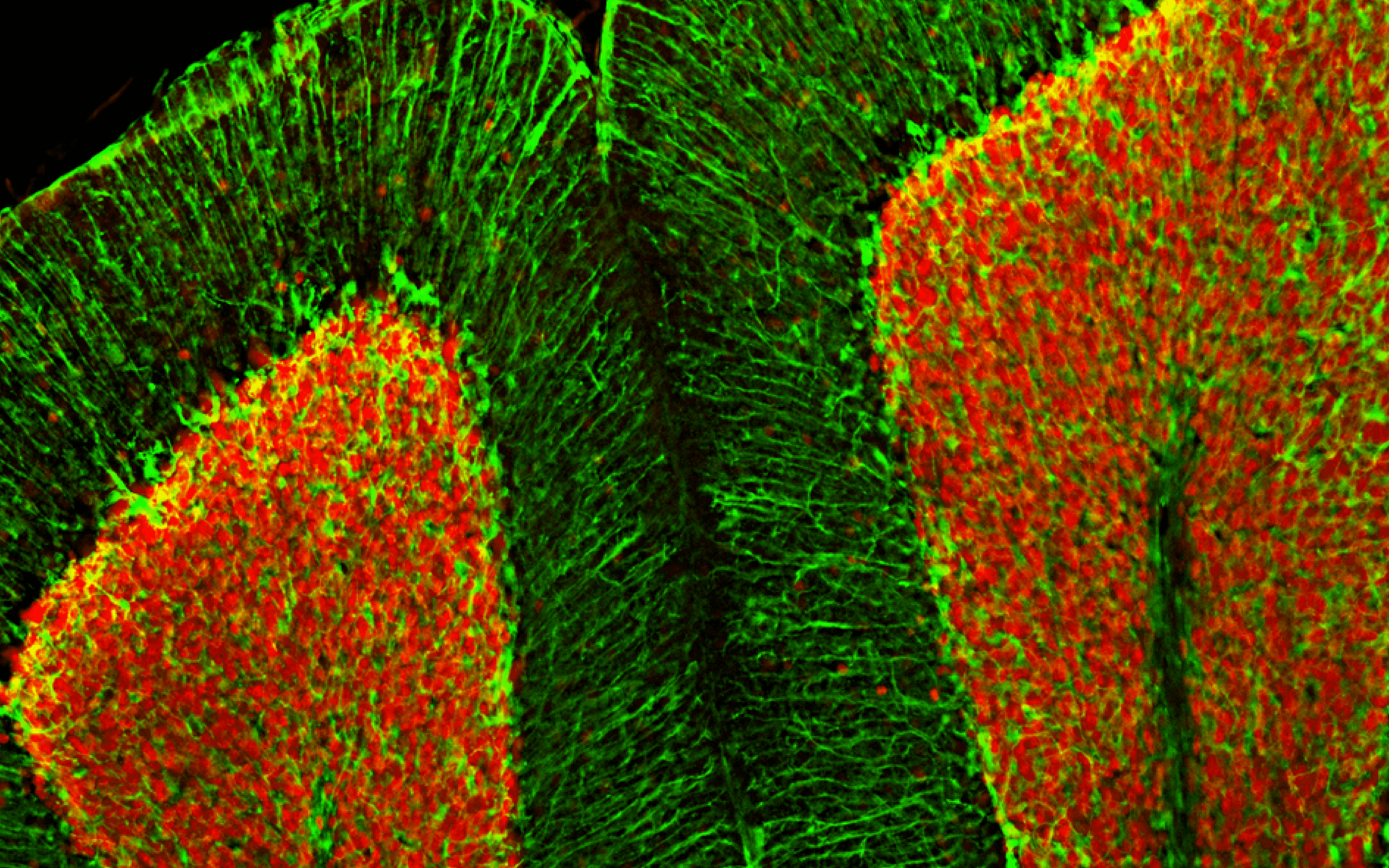
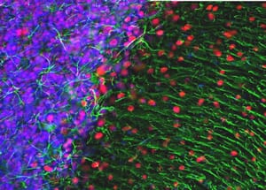
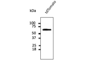
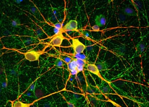
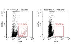
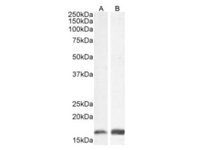
Primary Antibodies

Primary antibodies directly bind to specific antigens, with high specificity and affinity, for the purposes of purifying or detecting and measuring the antigens. Primary antibodies can either be developed as monoclonal antibodies, which bind to one specific epitope on the antigen, or polyclonal antibodies, which bind to multiple different epitopes on the antigen, using animals as the hosts, typically: rats, rabbits, mice, goats, and other species.

Primary antibodies can be raised against any antigen of research interest, including: proteins, peptides, carbohydrates, and other small molecules. Primary antibodies can also be raised to recognise post translational modifications such as phosphorylation, acetylation, methylation, and glycosylation. As such, primary antibodies are great tools to analyse components of living cells at the molecular level and identify proteins that may be involved in or may cause disease.

We offer over 65,000 unconjugated and directly conjugated primary antibodies directed against more than 14,000 targets. All antibodies are validated in multiple research applications and with samples derived from multiple different species. A growing number of antibodies have also been validated by protein array or undergone knockout validation to ensure specificity.

An antibody is a "Y-shaped" glycoprotein, produced by the body's immune system in response to invasion by foreign molecules, that is capable of binding to specific antigens. Each antibody is composed of four polypeptide chains, two identical heavy chains and two identical light chains, which vary in sequence and length. An antibody can be broken down into two F(ab) regions, the top sections of the "Y" which contain the variable region which binds specifically to a particular epitope on the antigen, and an Fc region, the bottom of the "Y" which provides a binding site for endogenous Fc receptors (and secondary antibodies).

Figure 1: Graphical representation of an antibody.

Antibody Graphic - Antibodies.com

Monoclonal antibodies are produced by injecting an antigen into a host animal in order to initiate a humoral immune response, and by subsequently extracting the antibody-producing spleen cells and fusing them in vitro with cultured malignant myeloma cells to create immortal hybridoma cell lines. This provides a stable, long-term supply of monoclonal antibodies. Monoclonal antibodies are identical, recognizing a single epitope, and are derived from a group of identical cloned cells. A clone ID is given to each clone of hybridoma cells which identifies the monoclonal antibody it produces.

Polyclonal antibodies are produced by injecting an antigen into a host animal in order to initiate a humoral immune response. After the initial immunisation, the host animal will receive a secondary, and potentially a tertiary, immunisation in order to produce higher concentrations of antibodies against the particular antigen. Polyclonal antibodies are a collection of multiple different antibodies, derived from the immune response of multiple B-cells, which each recognize a different epitope on the same antigen.

Monoclonal vs Polyclonal Antibodies

Polyclonal Monoclonal
A heterogeneous antibody population. A homogenous antibody population.
Lack of epitope specificity; will bind to multiple different epitopes on an antigen. High specificity and affinity for a single epitope on a single antigen.
Increased likelihood of cross-reactivity with similar antigens. Lower risk of cross-reactivity (due to specificity for a single epitope).
High batch-to-batch variability. No / low batch-to-batch variability.
Can create background noise in some applications. Create less background noise from staining of sections / cells.
Relatively inexpensive to produce. Significantly more expensive to produce.
Relatively quick to produce (~ 3 months). Slow to produce (~ 6 months).

In mammals, antibodies are classified into five main classes or isotypes according to the heavy chain they contain. These are: IgA (alpha), IgD (delta), IgE (epsilon), IgG (gamma), and IgM (mu). Each class differs in: the sequence of constant domains, the number of constant domains, the hinge structure, and the valency of the antibody.

The light chains of an antibody are classified as either kappa or lambda based on their polypeptide sequence. Typically, only one type of light chain is present in an individual antibody and, as such, the two light chains are identical.

Isotype Heavy Chain Light Chain Structure
IgG1 γ1 λ or κ Monomer
IgG2a γ2 λ or κ Monomer
IgG2b γ2 λ or κ Monomer
IgG3 γ3 λ or κ Monomer
IgG4 γ4 λ or κ Monomer
IgA1 α1 λ or κ Monomer or Dimer
IgA2 α2 λ or κ Monomer or Dimer
IgD δ λ or κ Monomer
IgE ε λ or κ Monomer
IgM μ λ or κ Pentamer

IgG (consisting of IgG1, IgG2a, IgG2b, IgG3, and IgG4) is the most abundant antibody in normal human serum, accounting for 70-85% of the total immunoglobulin pool. IgG is involved in placental transfer, with the mother’s IgG transferring to the foetus in order to provide immune protection during the initial stages of development.

Figure 2: Graphical representation of IgG.

IgG Isotype - Antibody Graphic - Antibodies.com

Each oval represents a protein domain in the antibody. These are systematically labelled by their chain type (L = Light chain or H = Heavy chain) and whether their sequence changes to bind an antigen (V = Variable domain or C = Constant domain), with CH domains numbered from the variable region. The domains are joined by linker regions of protein and disulfide bonds, represented by lines.

The antibody's structure is also described in terms of the fragments made by protein degradation or enzymatic cleavage of linker regions, namely the antigen-binding fragments (Fab), the variable fragments (Fv), and the constant fragment (Fc).

IgA (consisting of IgA1 and IgA2) accounts for around 5-15% of the antibody pool, existing either as a monomer or a dimer. IgA is the predominant antibody in mucous secretions, such as saliva, tears, and milk.

Figure 3: Graphical representation of IgA.

IgA Isotype - Antibody Graphic - Antibodies.com

IgD only accounts for approximately 1% of the total plasma immunoglobulin, however, it is found in large quantities on the membrane of B-cells. IgD has the same basic structure as IgG but has an extended hinge region, which is susceptible to proteolytic digestion. IgD is thought to serve as an antigen receptor for the activation of B cells.

Figure 4: Graphical representation of IgD.

IgD Isotype - Antibody Graphic - Antibodies.com

IgE only accounts for approximately 0.002% of serum antibodies and is found on the basophils and mast-cells. Similar to IgM, IgE has two additional constant domains instead of a hinge region. IgE is thought to play a role in immunity to parasites but is more commonly associated with type I immediate hypersensitivity, responding to innocuous environmental antigens such as pollen and peanuts, which can result in extreme reactions such as anaphylaxis or allergic rhinoconjunctivitis.

Figure 5: Graphical representation of IgE.

IgE Isotype - Antibody Graphic - Antibodies.com

IgM accounts for between 5-10% of the immunoglobulin population and is the predominant antibody in the body's primary immune response. It is often represented as a pentamer, with a five-chain structure held together by a J chain, however, it can also exist in a hexameric form, without the J chain, and as a monomer on the surface of B-cells. The large size of soluble IgM (~ 900kDa) mainly confines this antibody to the intravascular pool.

Figure 6: Graphical representation of IgM.

IgM Isotype - Antibody Graphic - Antibodies.com

Each clone number represents a specific cell line cloned from ascites that was used to manufacture the antibody. Since monoclonal antibodies are produced by more than one host, and more than one cell line, each cloned cell line receives a unique clone number to identify it.

Secondary antibodies should be raised against the host species of the primary antibody you are using. For example, if your primary antibody is a rat monoclonal you will require an anti-rat secondary antibody. We recommend checking the datasheet of the secondary antibody to ensure that it has been validated in the application you will be using.

Isotype controls are used to confirm that the binding of the primary antibody is specific and not a result of other protein interactions or non-specific Fc receptor binding.

The isotype control antibody should match the primary antibodies host species, isotype, and conjugation. For example, if the primary antibody is a HRP-conjugated rat IgG1, then you will need a HRP-conjugated rat IgG1 isotype control.

We recommend checking the datasheet first, which will often have a suggested positive control. It is important to ensure that the tissue or cell line used is from a tested species.

If no positive controls are suggested, we recommend looking at the UniProt website. This database often has a list of tissues that the protein is expressed in. These tissues can be considered suitable positive controls.

Many of our antibodies have dilution instructions on their datasheets. In these instances, we recommend following these instructions. For antibodies that do not have recommend dilutions, we recommend using the chart below to decide a starting dilution.

Application Tissue Culture Serum Ascites Whole Antiserum Purified Antibody
WB / DB 1/100 1/1,000 1/500 1 µg/ml
FC 1/100 1/1,000 1/500 1 µg/ml
ELISA 1/1,000 1/10,000 1/500 0.1 µg/ml
IHC / ICC Neat - 1/10 1/100 1/50 - 1/100 5 µg/ml
IP --- 1/100 1/50 - 1/100 1-10 µg/ml

Most unpurified antibodies (i.e. whole antiserum, culture supernatant, or ascites fluid) will not have a concentration stated on their datasheets, as it will not have been determined. These antibodies can vary significantly in specific antibody concentrations. As rough concentration estimates: tissue culture supernatants are 1-3 mg/ml, ascites are 5-10 mg/ml, and whole antisera are 1-10 mg/ml.

It is important to remember that the dilutions / concentrations in the chart above are recommended simply as a starting point. It may be necessary to adjust the dilution / concentration based on experimental results.

We recommend always storing the antibody as directed on the datasheet. We are unable to guarantee how the antibody will perform if it is stored under different conditions.

The size of the aliquots will depend on how much one typically uses in an experiment. The aliquots should be no smaller than 10µl. The smaller the aliquot is, the more the concentration is affected by evaporation and adsorption of the antibody onto the surface of the vial.

    Applications
    The research applications that a product has been tested to work in.
    Clonality
    The antibody is either monoclonal, binding to one specific epitope on the antigen, or polyclonal, binding to multiple different epitopes on the antigen.
    Clone ID
    A clone ID is given to an antibody produced by a single clone of hybridoma cells. Since antibodies are produced by more than one host or hybridoma line, each cloned cell line receives a unique clone number which identifies the monoclonal antibody it produces.
    Concentration
    The amount of antibody per amount of solution, as supplied. Usually expressed as mg/ml.
    Conjugate
    The fluorophore or reporter enzyme that the antibody is linked to. Unconjugated antibodies are not attached to a fluorophore or reporter enzyme and require further reagents, such as a secondary antibody, in order to visualise the antigen.
    Epitope
    An epitope is the antibody binding site on an antigen.
    Formulation
    The composition of the solution that the antibody is supplied in.
    Gene ID
    Gene ID is a database of gene specific content provided by the NIH.
    Host
    The species of animal that the antibody was raised in.
    Immunogen
    The immunogen is the specific type of antigen that was used to elicit the immune response in the host to generate the antibody. Also, commonly referred to as antigen.
    Isotype
    The structure of the antibody. Antibodies raised in mammals can have five different isotypes: IgG, IgM, IgA, IgD, and IgE. The isotype is based on the number of Y units and the type of heavy chain.
    KO Validated
    The antibody has been validated in a knockout cell line in order to confirm that it is specific for the intended antigen only.
    Product Form
    The antibody is either supplied as a liquid solution or lyophilised stock concentrates.
    Purification
    The method in which the antibody was purified.
    Purity
    The antibody may be supplied in a range of different purities; from crude antiserum and tissue culture supernatant to antigen-purified solutions.
    Reactivity
    The species in which the antibody has been shown to detect the antigen.
    Recommended Dilutions
    The dilutions recommended, as a starting point, for using a product in specific research applications. It may be necessary to adjust the dilution based on experimental results.
    Sequence
    The amino acid sequence of the immunogen used to raise the antibody.
    Specificity
    Each antibody is capable of binding to one unique epitope. The variable region, composed of 110-130 amino acids, gives the antibody its specificity for binding a particular position on the antigen.
    Storage
    The recommended storage instructions for the product in order to maintain its performance.
    Target
    The antigen that the antibody detects.
    Uniprot
    UniProt is a database of protein sequence and functional information.

Popular Primary Antibodies

Side Menu Main Menu Contact Us 0Checkout
Top