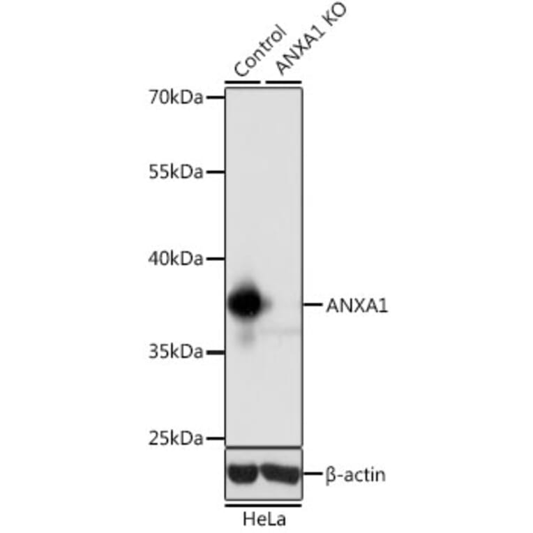
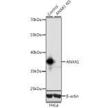
Western Blot - Anti-Annexin A1/ANXA1 Antibody (A13250)
Western blot analysis of extracts from normal (control) and ANXA1 knockout (KO) HeLa cells, using Anti-Annexin A1/ANXA1 Antibody (A13250) at 1:1,000 dilution. The secondary antibody was Goat Anti-Rabbit IgG H&L Antibody (HRP) at 1:10,000 dilution. Lysates/proteins were present at 25µg per lane. The blocking buffer used was 3% non-fat dry milk in TBST. Detection was with a ECL Basic Kit. Exposure time: 1s.
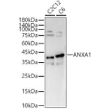
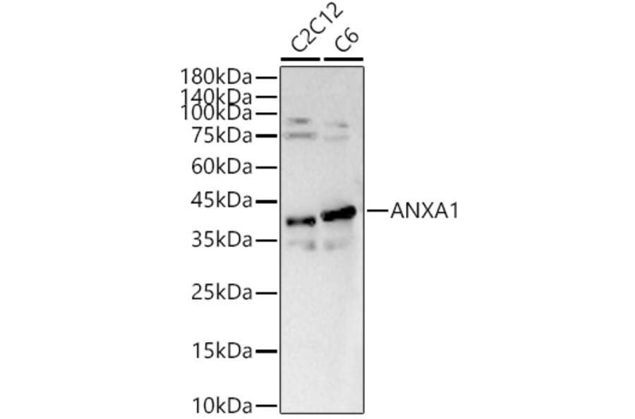
Western Blot - Anti-Annexin A1/ANXA1 Antibody (A13250)
Western blot analysis of various lysates, using Anti-Annexin A1/ANXA1 Antibody (A13250) at 1:700 dilution. The secondary antibody was Goat Anti-Rabbit IgG H&L Antibody (HRP) at 1:10,000 dilution. Lysates/proteins were present at 25µg per lane. The blocking buffer used was 3% non-fat dry milk in TBST. Detection was with a ECL Basic Kit. Exposure time: 90s.
Publishing research using Anti-Annexin A1/ANXA1 Antibody (A13250)? Please let us know so that we can list the citation on this page.