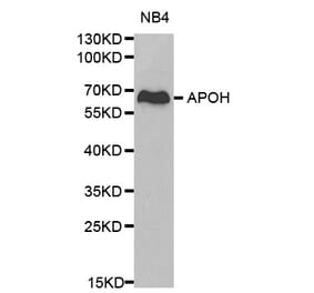
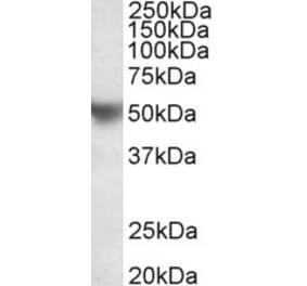
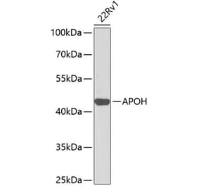
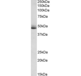
Unconjugated
Apolipoprotein (Apo) D is an important protein produced in many parts of the body. It is necessary for the development and repair of the brain and protection from oxidative stress. The purpose of this study was to investigate the extent to which apoD interacts with lipoproteins in human plasma. By using detergent-free ELISA, we show that immobilized monoclonal antibodies against apoD very efficiently bind to low density lipoprotein (LDL) from plasma; this binding is as equally efficient as binding to an anti-apoB monoclonal antibody. Adding detergent to the plasma inhibited the binding, suggesting that the binding is dependent on the presence of intact lipoprotein particles. Reversing the system by using immobilized anti-apoB revealed that the affinity of apoD for LDL is rather low, suggesting that multiple bindings are needed for a durable connection. Biosensor experiments using purified lipoproteins also showed that purified apoD and high density lipoprotein 3 (HDL3), a lipoprotein fraction rich in apoD, were both able to bind LDL very efficiently, indicating that the HDL3-LDL interaction may be a physiological consequence of the affinity of apoD for LDL. Furthermore, we found that apoD increases the binding of HDL to actively growing T24 bladder carcinoma cells but not to quiescent, contact-inhibited, confluent T24 cells. This result is especially intriguing given that the T24 supernatant only contained detectable levels of apoD after growth inhibition, raising the possibility that alternating the expression of apoD and a putative apoD-receptor could give direction to the flow of lipids. In the current paper, we conclude that apoD mediates binding of HDL to LDL and to growing T24 carcinomas, thereby highlighting the importance of apoD in lipid metabolism.
Background: Patients with antiphospholipid syndrome (APS) have a hypercoagulable condition associated with the presence of antiphospholipid autoantibodies (aPL). Consensus antibodies for diagnosis are lupus anticoagulant, anti-beta2 glycoprotein I (B2GPI) and anticardiolipin (IgG or IgM). Circulating immunocomplexes (CIC) of B2GPI associated with IgM or IgG were reported. Isolated IgA aB2GPI antibodies have achieved high diagnostic value although specific CIC of B2GPI bounded to IgA (B2A-CIC) has still not been described. CIC detection assays are mainly based on interaction with complement and are not appropriate to detect B2A-CIC because IgA does not fix complement using the classical pathway.
Patients and methods: Sera from healthy blood donors (N= 247) and from patients with thrombosis background and isolate positive for IgA aB2GPI (N = 68) were studied in a case-control study. Two methods were applied, these being a capture ELISA to quantify specific B2A-CIC and quantification of total IgA anti-B2GPI after dissociating CIC.
Results: B2A-CIC values in APS-patients were 19.27 ± 2.6 AU vs 6.1 ± 0.4 AU in blood donors (p < 0.001). There were 36.4% B2A-CIC positive patients (cutoff 21 AU) versus 5.5% in blood donors (p < 0.001). Dissociated IgA aB2GPI levels (total IgA aB2GPI) were 146.8 ± 10.8 IU/mL in patients vs. 22.4 IU/mL in controls (p < 0.001). B2A-CIC was independent of B2GPI and autoantibodies IgA aB2GPI serum levels.
Conclusion: B2A-CIC can be identified and quantified in an easy and reproducible manner using two complement-independent methods. The use of these tests in prospective studies will allow better understanding of the prognosis and outcome of patients.