- Primary Antibodies
- Secondary Antibodies
Fluorescent Conjugates
- Cyanine 3
- Cyanine 5
- Cyanine 5.5
- FITC
- PE
- Texas Red
- TRITC
- Unconjugated
Enzyme Conjugates
- Alkaline Phosphatase
- Biotin
- HRP
Application
- ELISA
- ICC/IF
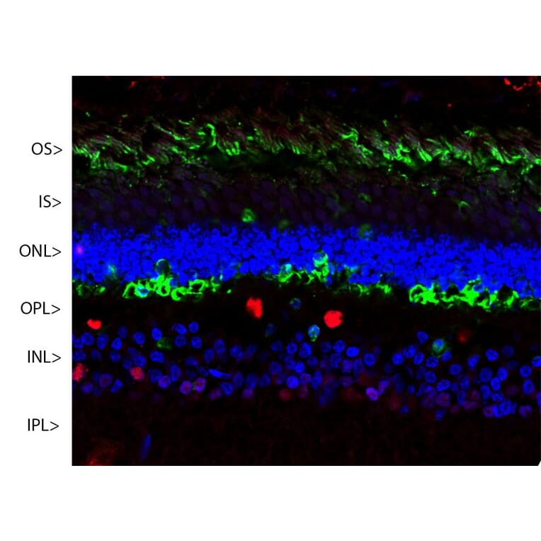
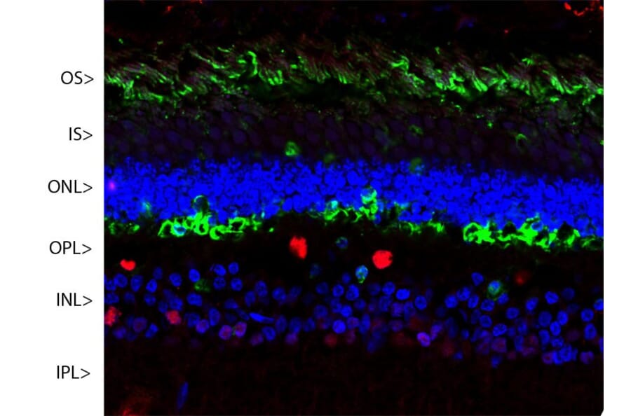
- IHC
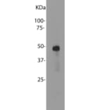
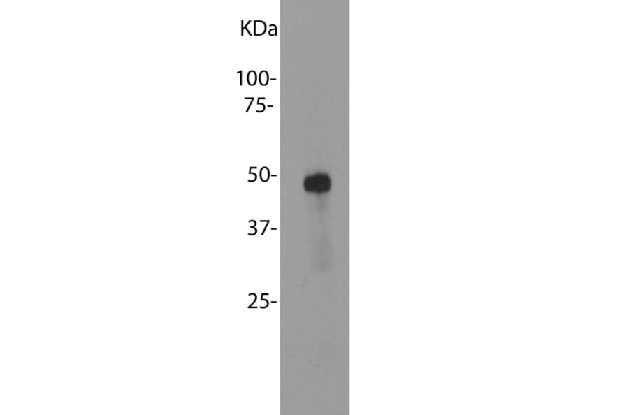
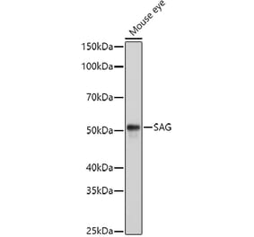
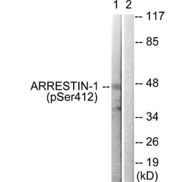
- Western Blot
- Proteins & Peptides
- ELISA Kits
- Custom Services
- Research Areas
- Customer SupportSupport