Immunocytochemistry/Immunofluorescence analysis of human neuroblastoma cell line (SK-N-BE, fixed in 4% formaldehyde for 15 min at room temperature, using Anti-Ataxin 1 Antibody [S76-8] (A305028), at 1:100 for 60 minutes at room temperature. The secondary antibody used was Goat Anti-Mouse ATTO 488 at 1:200 for 60 minutes at room temperature. Counterstain: Phalloidin Texas Red F-Actin stain; DAPI (blue) nuclear stain at 1:1000, 1:5,000 for 60 minutes at room temperature, 5 minutes at room temperature. Localization: Cytoplasm, Nucleus. Magnification: 60X.(A) DAPI (blue) nuclear stain. (B) Phalloidin Texas Red F-Actin stain. (C) Ataxin 1 Antibody. (D) Composite.
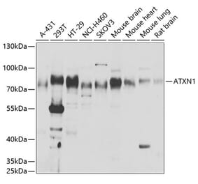
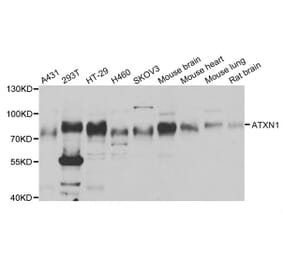
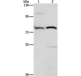
Figure 2: Western Blot - Anti-Ataxin 1 Antibody [S76-8] (A305028)
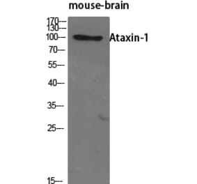
Western blot analysis of Monkey COS-1 cells transfected with Ataxin- 1 showing detection of ~85 kDa Ataxin 1 protein using Anti-Ataxin 1 Antibody [S76-8] (A305028) at 1:200 for 16 hours at 4°C. Lane 1: Molecular Weight Ladder. Lane 2: Monkey COS-1 cells transfected with Ataxin- 1. Load: 15 µg. Block: 2% BSA and 2% Skim Milk in 1X TBST. The secondary antibody used was Goat Anti-Mouse IgG: HRP at 1:1,000 for 1 hour room temperature. Color Development: ECL solution for 6 min in room temperature. Predicted/Observed Size: ~85 kDa.
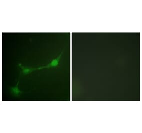
Immunocytochemistry/Immunofluorescence analysis of human neuroblastoma cells (SH-SY5Y), fixed in 4% PFA for 15 min, using Anti-Ataxin 1 Antibody [S76-8] (A305028), at 1:100 for overnight at 4°C with slow rocking. The secondary antibody used was AlexaFluor 488 at 1:1,000 for 1 hour at room temperature. Counterstain: Phalloidin-iFluor 647 (red) F-Actin stain; Hoechst (blue) nuclear stain at 1:800, 1.6mM for 20 minutes at room temperature.(A) Hoechst (blue) nuclear stain. (B) Phalloidin-iFluor 647 (red) F-Actin stain. (C) Ataxin 1 Antibody (D) Composite.
Publishing research using Anti-Ataxin 1 Antibody [S76-8] (A305028)? Please let us know so that we can list the citation on this page.
Alternative products to Anti-Ataxin 1 Antibody [S76-8] (A305028)


![Immunocytochemistry/Immunofluorescence - Anti-Ataxin 1 Antibody [S76-8] (A305028) - Antibodies.com](https://cdn.antibodies.com/image/catalog/305/A305028_1.png?profile=product_top)
![Western Blot - Anti-Ataxin 1 Antibody [S76-8] (A305028) - Antibodies.com](https://cdn.antibodies.com/image/catalog/305/A305028_2.png?profile=product_top)
![Immunocytochemistry/Immunofluorescence - Anti-Ataxin 1 Antibody [S76-8] (A305028) - Antibodies.com](https://cdn.antibodies.com/image/catalog/305/A305028_3.png?profile=product_top)
![Immunocytochemistry/Immunofluorescence - Anti-Ataxin 1 Antibody [S76-8] (A305028) - Antibodies.com](https://cdn.antibodies.com/image/catalog/305/A305028_1.png?profile=product_top_thumb)
![Western Blot - Anti-Ataxin 1 Antibody [S76-8] (A305028) - Antibodies.com](https://cdn.antibodies.com/image/catalog/305/A305028_2.png?profile=product_top_thumb)
![Immunocytochemistry/Immunofluorescence - Anti-Ataxin 1 Antibody [S76-8] (A305028) - Antibodies.com](https://cdn.antibodies.com/image/catalog/305/A305028_3.png?profile=product_top_thumb)
![Immunocytochemistry/Immunofluorescence - Anti-Ataxin 1 Antibody [S76-8] (A305028) - Antibodies.com](https://cdn.antibodies.com/image/catalog/305/A305028_1.png?profile=product_image)
![Western Blot - Anti-Ataxin 1 Antibody [S76-8] (A305028) - Antibodies.com](https://cdn.antibodies.com/image/catalog/305/A305028_2.png?profile=product_image)
![Immunocytochemistry/Immunofluorescence - Anti-Ataxin 1 Antibody [S76-8] (A305028) - Antibodies.com](https://cdn.antibodies.com/image/catalog/305/A305028_3.png?profile=product_image)
![Western Blot - Anti-Ataxin 1 Antibody [S65-37] (A304806) - Antibodies.com](https://cdn.antibodies.com/image/catalog/304/A304806_1.png?profile=product_alternative)