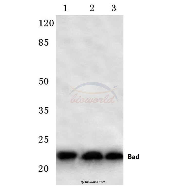
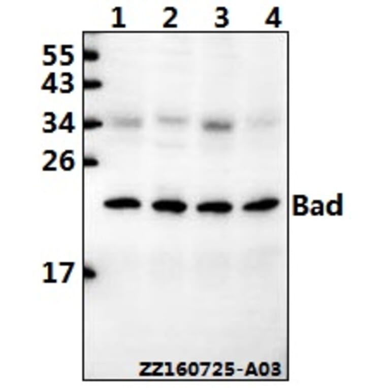
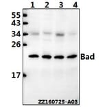
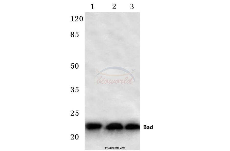
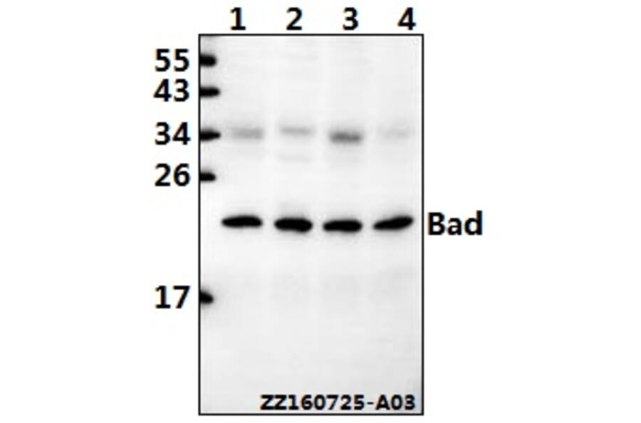
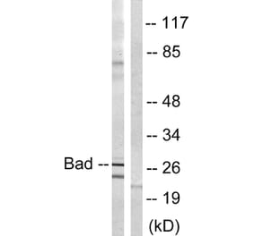
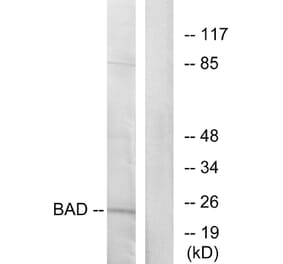
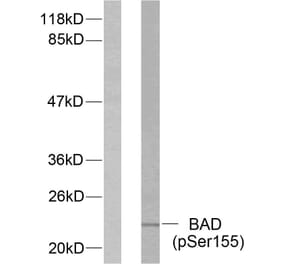
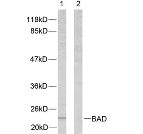
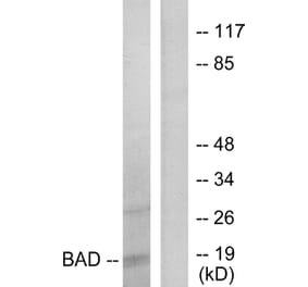
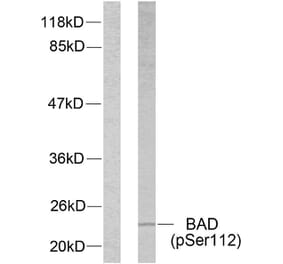
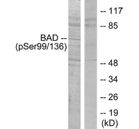
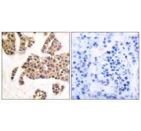
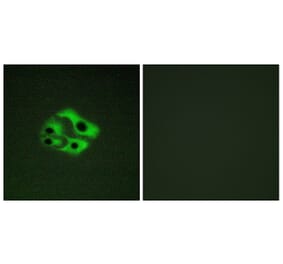
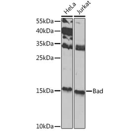
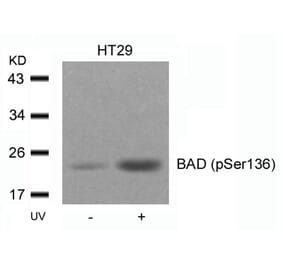
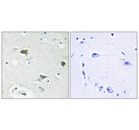
Bad (F130) pAb detects endogenous levels of Bad protein.
Applications
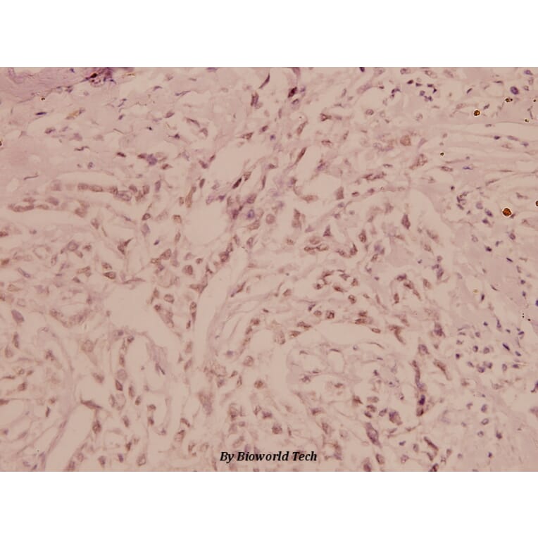
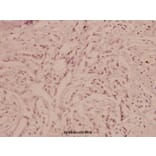
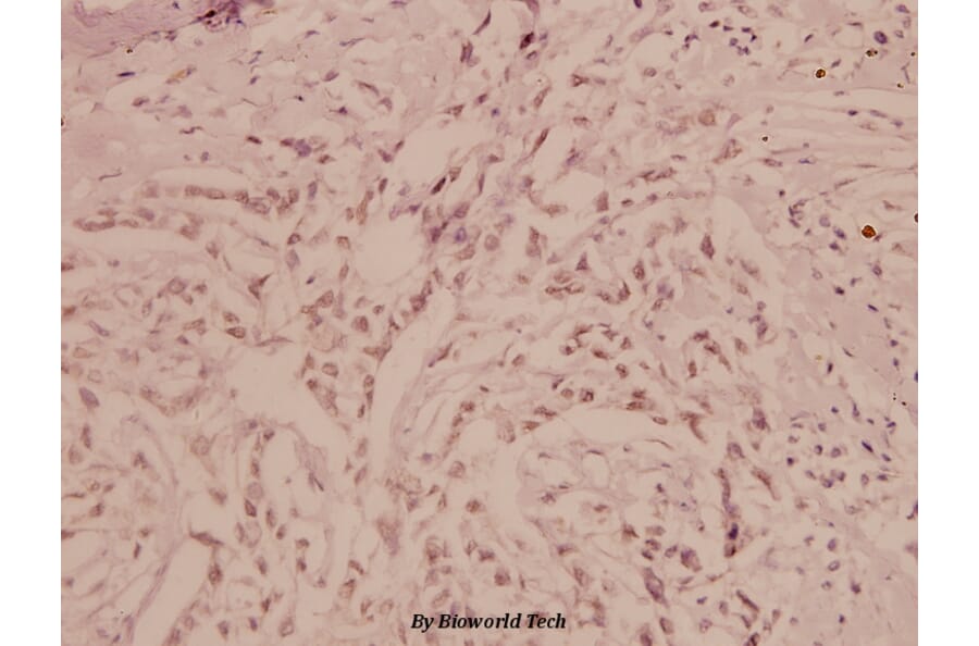
WB, IHC
Reactivity
Human, Mouse, Rat
Immunogen
Synthetic peptide, corresponding to amino acids 100-150 of Human Bad.
Host
Rabbit
Clonality
Polyclonal
Conjugate
Unconjugated
Molecular Weight
~ 24 kDa
Purity
The antibody was affinity-purified from rabbit antiserum by affinity-chromatography using epitope-specific immunogen and the purity is > 95% (by SDS-PAGE).
Product Form
1 mg/ml in Phosphate buffered saline (PBS) with 0.05% sodium azide, approx. pH 7.2.
Synonyms
BBC6, Bcl-2-binding component 6, Bcl-2-like protein 8, Bcl-xL/Bcl-2-associated death promoter, Bcl2 antagonist of cell death, Bcl2-associated agonist of cell death, Bcl2-L-8, BCL2L8