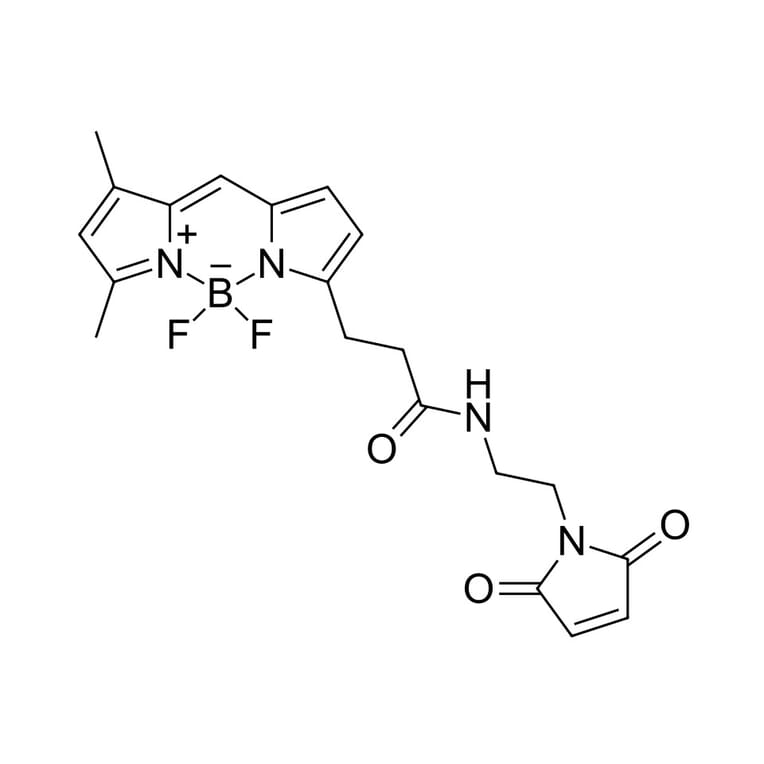
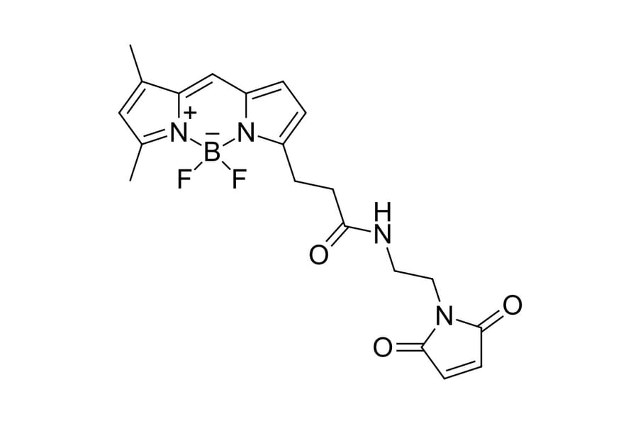
Many discoveries in cell biology rely on making specific proteins visible within their native cellular environment. There are various genetically encoded tags, such as fluorescent proteins, developed for fluorescence microscopy (FM). However, there are almost no genetically encoded tags that enable cellular proteins to be observed by both FM and electron microscopy (EM). Herein, we describe a technology for labeling proteins with diverse chemical reporters, including bright organic fluorophores for FM and electron-dense nanoparticles for EM. Our technology uses versatile interacting peptide (VIP) tags, a class of genetically encoded tag. We present VIPER, which consists of a coiled-coil heterodimer formed between the genetic tag, CoilE, and a probe-labeled peptide, CoilR. Using confocal FM, we demonstrate that VIPER can be used to highlight subcellular structures or to image receptor-mediated iron uptake. Additionally, we used VIPER to image the iron uptake machinery by correlative light and EM (CLEM). VIPER compared favorably with immunolabeling for imaging proteins by CLEM, and is an enabling technology for protein targets that cannot be immunolabeled. VIPER is a versatile peptide tag that can be used to label and track proteins with diverse chemical reporters observable by both FM and EM instrumentation.
Protein translocation and insertion into the bacterial cytoplasmic membrane are the essential processes mediated by the Sec machinery. The core machinery is composed of the membrane-embedded translocon SecYEG that interacts with the secretion-dedicated ATPase SecA and translating ribosomes. Despite the simplicity and the available structural insights on the system, diverse molecular mechanisms and functional dynamics have been proposed. Here, we employ total internal reflection fluorescence microscopy to study the oligomeric state and diffusion of SecYEG translocons in supported lipid bilayers at the single-molecule level. Silane-based coating ensured the mobility of lipids and reconstituted translocons within the bilayer. Brightness analysis suggested that approx. 70% of the translocons were monomeric. The translocons remained in a monomeric form upon ribosome binding, but partial oligomerization occurred in the presence of nucleotide-free SecA. Individual trajectories of SecYEG in the lipid bilayer revealed dynamic heterogeneity of diffusion, as translocons commonly switched between slow and fast mobility modes with corresponding diffusion coefficients of 0.03 and 0.7 µm2 ·s-1 . Interactions with SecA ATPase had a minor effect on the lateral mobility, while bound ribosome:nascent chain complexes substantially hindered the diffusion of single translocons. Notably, the mobility of the translocon:ribosome complexes was not affected by the solvent viscosity or macromolecular crowding modulated by Ficoll PM 70, so it was largely determined by interactions within the lipid bilayer and at the interface. We suggest that the complex mobility of SecYEG arises from the conformational dynamics of the translocon and protein:lipid interactions.