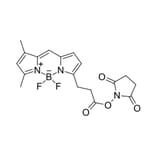
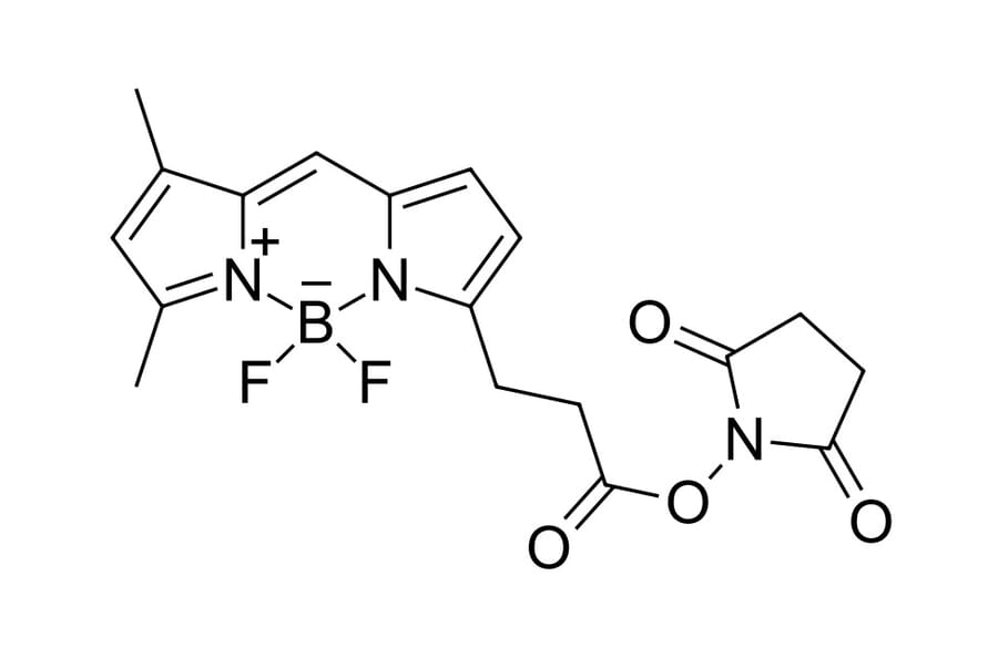
The vertiginous increase in the use of extracellular vesicles and especially exosomes for therapeutic applications highlights the necessity of advanced techniques for gaining a deeper knowledge of their pharmacological properties. Herein, we report a novel chemical approach for the robust attachment of commercial fluorescent dyes to the exosome surface with covalent binding. The applicability of the methodology was tested on milk and cancer cell-derived exosomes (from U87 and B16F10 cancer cells). We demonstrated that fluorescent labeling did not modify the original physicochemical properties of exosomes. We tested this nanoprobe in cell cultures and healthy mice to validate its use for in vitro and in vivo applications. We confirmed that these fluorescently labeled exosomes could be successfully visualized with optical imaging.
Silica nanoparticles (NPs) are widely used in various industrial and biomedical applications. Little is known about the cellular uptake of co-exposed silica particles, as can be expected in our daily life. In addition, an inflamed microenvironment might affect a NP's uptake and a cell's physiological response. Herein, prestimulated mouse J774A.1 macrophages with bacterial lipopolysaccharide were post-exposed to micron- and nanosized silica particles, either alone or together, i.e., simultaneously or sequentially, for different time points. The results indicated a morphological change and increased expression of tumor necrosis factor alpha in lipopolysaccharide prestimulated cells, suggesting a M1-polarization phenotype. Confocal laser scanning microscopy revealed the intracellular accumulation and uptake of both particle types for all exposure conditions. A flow cytometry analysis showed an increased particle uptake in lipopolysaccharide prestimulated macrophages. However, no differences were observed in particle uptakes between single- and co-exposure conditions. We did not observe any colocalization between the two silica (SiO2) particles. However, there was a positive colocalization between lysosomes and nanosized silica but only a few colocalized events with micro-sized silica particles. This suggests differential intracellular localizations of silica particles in macrophages and a possible activation of distinct endocytic pathways. The results demonstrate that the cellular uptake of NPs is modulated in inflamed macrophages but not in the presence of micron-sized particles.