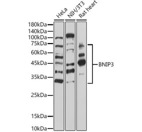
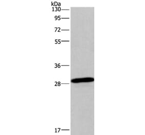
Figure 1: Western Blot - Anti-BNIP3 Antibody [ARC50531] (A305317)
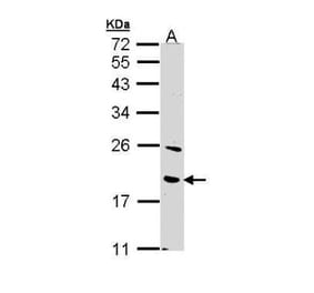
Western blot analysis of extracts of various cell lines, using Anti-BNIP3 Antibody [ARC50531] (A305317) at 1:1,000 dilution. Hela cells and NIH/3T3 cells were treated by Cobalt chloride (0. 1 mM) at 37°C for 4 hours. The secondary antibody was Goat Anti-Rabbit IgG H&L Antibody (HRP) at 1:10,000 dilution. Lysates/proteins were present at 25µg per lane. The blocking buffer used was 3% non-fat dry milk in TBST. Detection was with a ECL Basic Kit. Exposure time: 30s.
Immunohistochemistry analysis of paraffin-embedded human cervix cancer tissue using Anti-BNIP3 Antibody [ARC50531] (A305317) at a dilution of 1:500 (40x lens). Perform high pressure antigen retrieval with 10 mM citrate buffer pH 6.0 before commencing with IHC staining protocol.
Immunohistochemistry analysis of paraffin-embedded human liver using Anti-BNIP3 Antibody [ARC50531] (A305317) at a dilution of 1:500 (40x lens). Perform high pressure antigen retrieval with 10 mM citrate buffer pH 6.0 before commencing with IHC staining protocol.
Immunohistochemistry analysis of paraffin-embedded rat heart using Anti-BNIP3 Antibody [ARC50531] (A305317) at a dilution of 1:500 (40x lens). Perform high pressure antigen retrieval with 10 mM citrate buffer pH 6.0 before commencing with IHC staining protocol.
Immunohistochemistry analysis of paraffin-embedded rat kidney using Anti-BNIP3 Antibody [ARC50531] (A305317) at a dilution of 1:500 (40x lens). Perform high pressure antigen retrieval with 10 mM citrate buffer pH 6.0 before commencing with IHC staining protocol.


![Western Blot - Anti-BNIP3 Antibody [ARC50531] (A305317) - Antibodies.com](https://cdn.antibodies.com/image/catalog/305/A305317_1.jpg?profile=product_top)
![Immunohistochemistry - Anti-BNIP3 Antibody [ARC50531] (A305317) - Antibodies.com](https://cdn.antibodies.com/image/catalog/305/A305317_2.jpg?profile=product_top)
![Immunohistochemistry - Anti-BNIP3 Antibody [ARC50531] (A305317) - Antibodies.com](https://cdn.antibodies.com/image/catalog/305/A305317_3.jpg?profile=product_top)
![Immunohistochemistry - Anti-BNIP3 Antibody [ARC50531] (A305317) - Antibodies.com](https://cdn.antibodies.com/image/catalog/305/A305317_4.jpg?profile=product_top)
![Immunohistochemistry - Anti-BNIP3 Antibody [ARC50531] (A305317) - Antibodies.com](https://cdn.antibodies.com/image/catalog/305/A305317_5.jpg?profile=product_top)
![Western Blot - Anti-BNIP3 Antibody [ARC50531] (A305317) - Antibodies.com](https://cdn.antibodies.com/image/catalog/305/A305317_1.jpg?profile=product_top_thumb)
![Immunohistochemistry - Anti-BNIP3 Antibody [ARC50531] (A305317) - Antibodies.com](https://cdn.antibodies.com/image/catalog/305/A305317_2.jpg?profile=product_top_thumb)
![Immunohistochemistry - Anti-BNIP3 Antibody [ARC50531] (A305317) - Antibodies.com](https://cdn.antibodies.com/image/catalog/305/A305317_3.jpg?profile=product_top_thumb)
![Immunohistochemistry - Anti-BNIP3 Antibody [ARC50531] (A305317) - Antibodies.com](https://cdn.antibodies.com/image/catalog/305/A305317_4.jpg?profile=product_top_thumb)
![Immunohistochemistry - Anti-BNIP3 Antibody [ARC50531] (A305317) - Antibodies.com](https://cdn.antibodies.com/image/catalog/305/A305317_5.jpg?profile=product_top_thumb)
![Western Blot - Anti-BNIP3 Antibody [ARC50531] (A305317) - Antibodies.com](https://cdn.antibodies.com/image/catalog/305/A305317_1.jpg?profile=product_image)
![Immunohistochemistry - Anti-BNIP3 Antibody [ARC50531] (A305317) - Antibodies.com](https://cdn.antibodies.com/image/catalog/305/A305317_2.jpg?profile=product_image)
![Immunohistochemistry - Anti-BNIP3 Antibody [ARC50531] (A305317) - Antibodies.com](https://cdn.antibodies.com/image/catalog/305/A305317_3.jpg?profile=product_image)
![Immunohistochemistry - Anti-BNIP3 Antibody [ARC50531] (A305317) - Antibodies.com](https://cdn.antibodies.com/image/catalog/305/A305317_4.jpg?profile=product_image)
![Immunohistochemistry - Anti-BNIP3 Antibody [ARC50531] (A305317) - Antibodies.com](https://cdn.antibodies.com/image/catalog/305/A305317_5.jpg?profile=product_image)