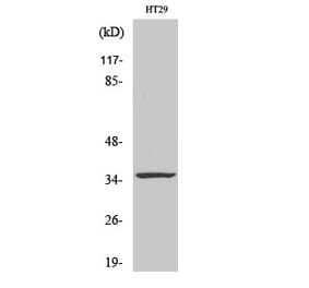
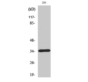
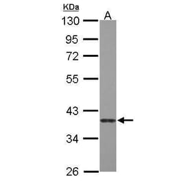
Anti-Caspase 7 (M45) Antibody (A26960) has been discontinued and is no longer available.
View all Anti-Caspase 7 Antibodies.
Unconjugated
Recent studies have reported that caspase 7 has an apoptotic and nonapoptotic function. However, the relationship between caspase 7 and spermatogenesis remains unknown. This study aimed to investigate the possible function of caspase 7 during normal and abnormal spermatogenesis. The cleaved form of caspase 7 was detected in testis tissues at different postpartum times (5-14 weeks) by qRT-PCR, Western blot and immunohistochemistry (IHC). Then, the mice models of spermatogenic dysfunction were obtained by busulfan (30 mg kg-1 to further evaluate the potential function and mechanism of caspase 7. qRT-PCR and Western blot results showed that caspase 7 expression was gradually elevated from 5 to 14 weeks, which was not connected with apoptosis. IHC results revealed that caspase 7 was mainly located in spermatogenic cells and Leydig cells. In addition, spermatogenic dysfunction induced by busulfan gradually enhanced the apoptosis and elevated the expression of caspase 3, caspase 6, and caspase 9, but decreased the expression of caspase 7 in spermatogenic cells. However, when spermatogenic cells were mostly disappeared at the fourth week after busulfan treatment, caspase 7 expression in Leydig cells was significantly increased and positively correlated with the expression of caspase 3, caspase 6, and caspase 9. Therefore, these results indicate that caspase 7 has a nonapoptic function that participates in normal spermatogenesis, but also displays apoptotic function in spermatogenic dysfunction.
Our group was the first one reporting that autophagy could be triggered by airborne fine particulate matter (PM) with a mean diameter of less than 2.5 μm (PM2.5) in human lung epithelial A549 cells, which could potentially lead to cell death. In the present study, we further explored the potential interactions between autophagy and apoptosis because it was well documented that PM2.5 could induce apoptosis in A549 cells. Much to our surprise, we found that PM2.5-exposure caused oxidative stress, resulting in activation of multiple cell death pathways in A549 cells, that is, the tumor necrosis factor-alpha (TNF-α)-induced pathway as evidenced by TNF-α secretion and activation of caspase-8 and -3, the intrinsic apoptosis pathway as evidenced by increased expression of pro-apoptotic protein Bax, decreased expression of anti-apoptotic protein Bcl-2, disruption of mitochondrial membrane potential, and activation of caspase-9 and -3, and autophagy as evidenced by an increased number of double-membrane vesicles, accompanied by increases of conversion and punctuation of microtubule-associated proteins light chain 3 (LC3) and expression of Beclin 1. It appears that reactive oxygen species (ROS) function as signaling molecules for all the three pathways because pretreatment with N-acetylcysteine, a scavenger of ROS, almost completely abolished TNF-α secretion and significantly reduced the number of apoptotic and autophagic cells. In another aspect, inhibiting autophagy with 3-methyladenine, a specific autophagy inhibitor, enhanced PM2.5-induced apoptosis and cytotoxicity. Intriguingly, neutralization of TNF-α with an anti-TNF-α special antibody not only abolished activation of caspase-8, but also drastically reduced LC3-II conversion. Thus, the present study has provided novel insights into the mechanism of cytotoxicity and even pathogenesis of diseases associated with PM2.5 exposure.