Unconjugated
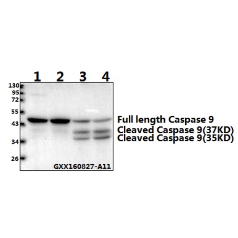
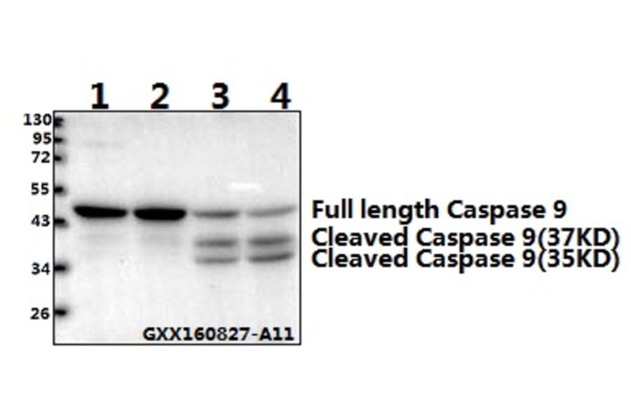
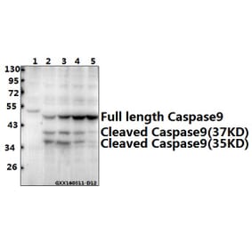
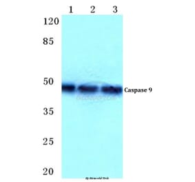
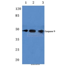
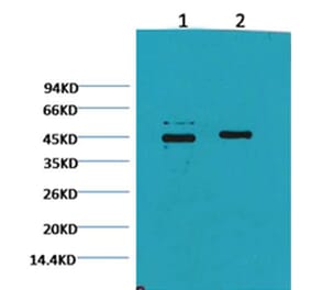
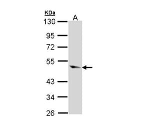
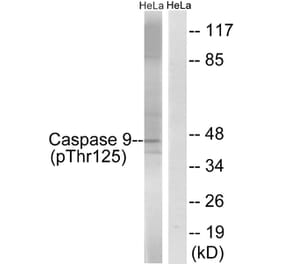
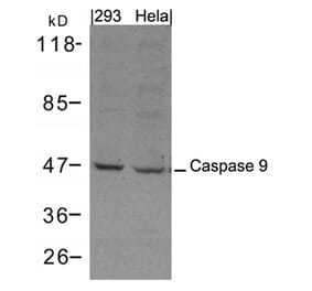
Cyclin D2 is involved in the pathology of vascular complications of type 2 diabetes mellitus (T2DM). This study investigated the role of cyclin-D2-regulated miRNAs in endothelial cell proliferation of T2DM. Results showed that higher glucose concentration (4.5 g/l) significantly promoted the proliferation of rat aortic endothelial cells (RAOECs), and significantly increased the expression of cyclin D2 and phosphorylation of retinoblastoma 1 (p-RB1) in RAOECs compared with those under low glucose concentration. The cyclin D2-3' untranslated region is targeted by miR-98, as demonstrated by miRNA analysis software. Western blot also confirmed that cyclin D2 and p-RB1 expression was regulated by miR-98. The results indicated that miR-98 treatment can induce RAOEC apoptosis. The suppression of RAOEC growth by miR-98 might be related to regulation of Bcl-2, Bax and Caspase 9 expression. Furthermore, the expression levels of miR-98 decreased in 4.5 g/l glucose-treated cells compared with those treated by low glucose concentration. Similarly, the expression of miR-98 significantly decreased in aortas of established streptozotocin (STZ)-induced diabetic rat model compared with that in control rats; but cyclin D2 and p-RB1 levels remarkably increased in aortas of STZ-induced diabetic rats compared with those in healthy control rats. In conclusion, this study demonstrated that high glucose concentration induces cyclin D2 up-regulation and miR-98 down-regulation in the RAOECs. By regulating cyclin D2, miR-98 can inhibit human endothelial cell growth, thereby providing novel therapeutic targets for vascular complication of T2DM.
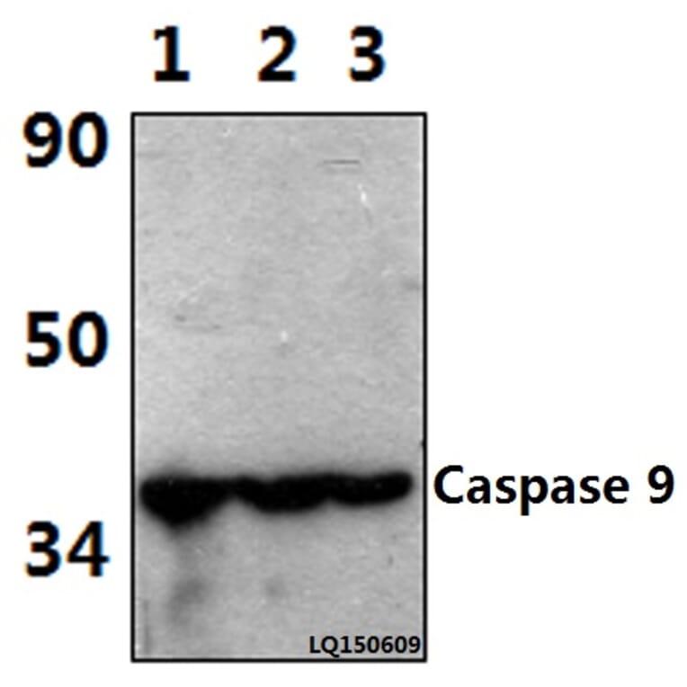
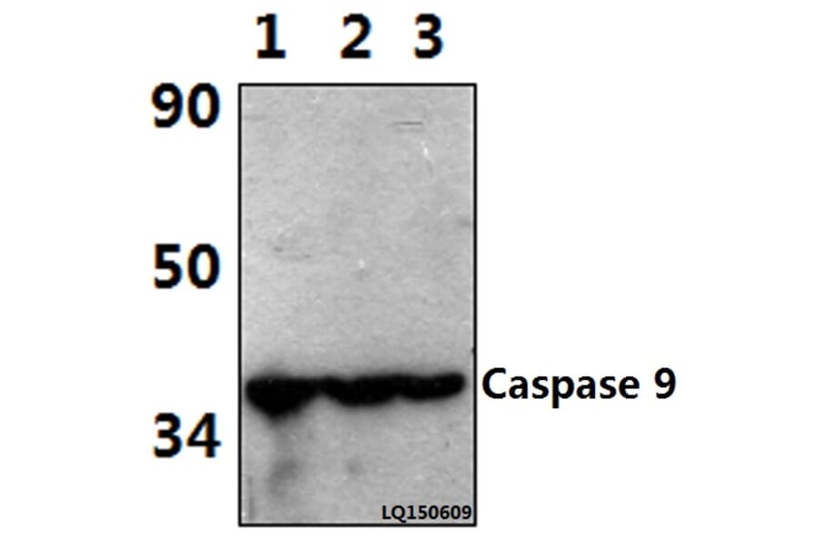
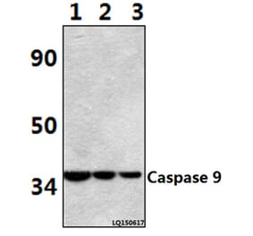
Radiotherapy is a widespread treatment in human solid tumors. However, therapy resistance and poor prognosis are still problems. Gambogic acid (GA), extracted from the dried yellow resin of gamboges, has an anticancer effect against various types of cancer cells. To explore the radiosensitivity of GA on esophageal cancer cell line TE13, cell viability was tested by Cell Counting Kit-8 (CCK-8) assay, colony formation assay was used to assess the effects of GA on the radiosensitivity of TE13, and flow cytometry was performed to meter the percentage of apoptosis. The protein levels of microtubule-associated protein 1 light chain 3 (LC3), caspase3, caspase8, casepase9, pAkt, and p-mammalian target of rapamycin (p-mTOR) were tested using Western blot. The distribution of LC3 was detected by immunofluorescence. Additionally, we also examined reactive oxygen species (ROS) expression by laser scanning confocal microscope (LSCM). The cells were transfected with adenovial vector to monitor the autophagy through the expression of green fluorescent protein (GFP-red fluroscent protein (RFP)-LC3. The rates of apoptotic cells in combined-treated TE13 increased significantly compared with the control groups in accordance with the results of Western blot. The clonogenic survival assay showed that GA enhances radiosensitivity with a sensitizing enhancement ratio (SER) of 1.217 and 1.436 at different concentrations. The LC3-II protein level increased in the combined group indicating the increase of autophagy incidence, and the results of GFP-RFP-LC3 experiment showed that GA may block the process of autophagic flux in TE13 cells. Moreover, we successfully demonstrated that ROS is involved in the induction of autophagy. ROS-mediated autophagy depends on the inhibition of the Akt/mTOR pathway. Taken together, GA induced radiosensitivity involves autophagy and apoptosis which are regulated by ROS hypergeneration and Akt/mTOR inhibition.