ISO 9001:2015 Certified
Live Customer Support
4.5/5 on Trustpilot
100% Quality Guarantee

mIHC Lung Cancer TME Panel I - 5 Color (A334689)

$1,537

Shipping Information

$40
Dispatched from St. Louis, MO.
Lead Time: 7-10 business days.
Name
mIHC Lung Cancer TME Panel I - 5 Color
Description
Ready-to-use 5-plex panel for lung cancer tumor microenvironment, includes PD-L1, PD 1, Ki67, SOX2, GSTP1 [520, 570, 620, 650, 700].
Applications
IHC, mIHC, TMA
Recommended Dilutions
The fluorescent dye is provided as a 100× stock solution. To prepare the working solution, dilute the stock 1:100 with the freshly prepared signal amplification reaction solution. DAPI should be diluted 1:100 in sterile water to obtain its working solution. The kit also contains a universal secondary antibody compatible with mouse and rabbit primary antibodies; before use, verify that the primary antibody species matches the secondary antibody. The secondary antibody is supplied as a 1× working solution and is ready for use.
Principle of Assay
This multiplex fluorescence immunohistochemical assay is based on the specific antigen-antibody interaction combined with tyramide signal amplification (TSA) technology. Similar to the DAB chromogenic reaction used in conventional IHC, TSA employs horseradish peroxidase (HRP)-conjugated secondary antibodies to catalyze the activation of a tyramide-linked fluorophore substrate. The activated fluorophore covalently binds to tyrosine residues in proximity to the target antigen, generating a stable and permanent fluorescent signal. Following each staining cycle, non-covalently bound antibodies are removed through heat-mediated antigen retrieval, enabling subsequent rounds of staining with different primary antibodies and fluorophores. Through iterative cycles, multiple biomarkers, such as PD-L1, PD 1, Ki67, SOX2, and GSTP1, can be sequentially labeled within the same lung cancer tissue section. This approach allows high-resolution visualization of spatial co-expression patterns and cellular interactions within the tumor microenvironment, providing a comprehensive molecular and cellular profile critical for diagnostic and translational research.
Sample Type
Lung cancer.
Components
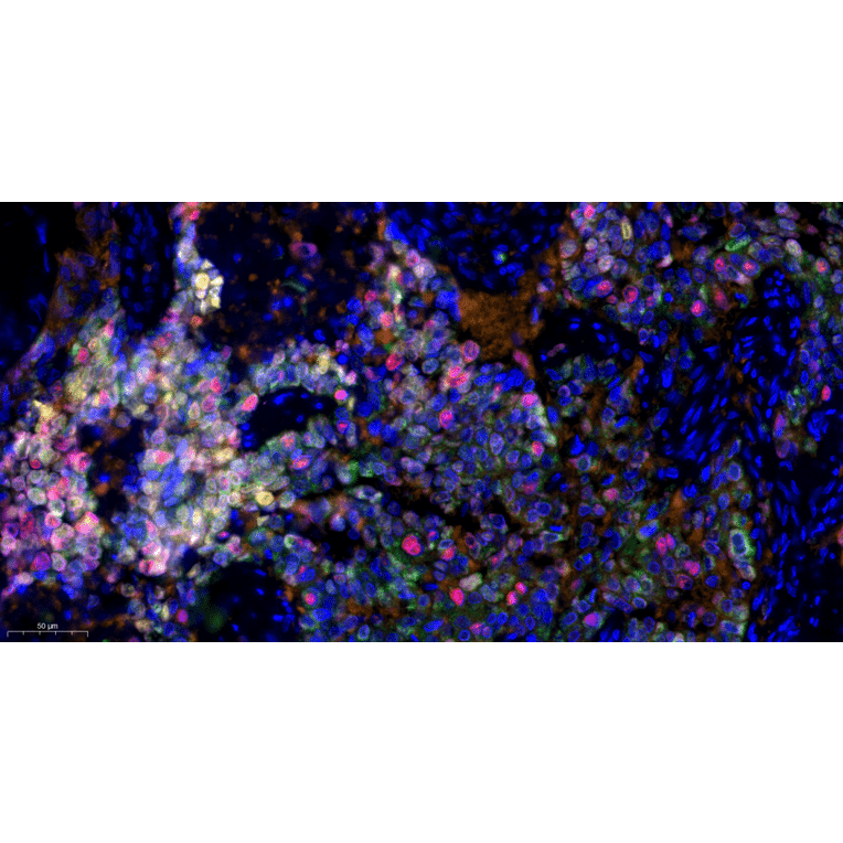
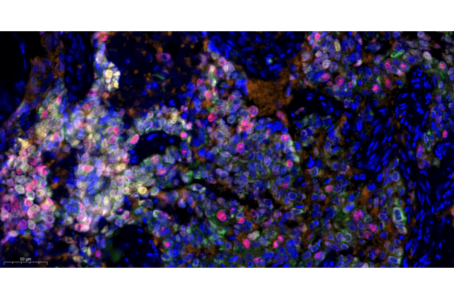
The kit contains PD-L1 rabbit monoclonal antibody, PD 1 rabbit monoclonal antibody, Ki67 rabbit monoclonal antibody, SOX2 rabbit monoclonal antibody, and GSTP1 rabbit monoclonal antibody, which are used to specifically recognize their respective protein targets in tissue or cell samples. It also includes TSA monochromatic fluorescent dye 520, TSA monochromatic fluorescent dye 570, TSA monochromatic fluorescent dye 620, TSA monochromatic fluorescent dye 650, and TSA monochromatic fluorescent dye 700. These dyes emit green, yellow-orange, red, far-red, and near-infrared light respectively and are used with tyramide signal amplification to visualize multiple targets in the same sample. The kit contains a signal amplification reaction solution, which provides the reagents necessary for the TSA reaction to deposit the fluorescent dye at the site of the target. A Pika universal HRP-labeled secondary antibody is included, which binds to the primary antibodies and catalyzes the TSA reaction. The kit also contains an anti-fluorescence quenching tablet to protect the fluorescent signal from fading during imaging. Finally, DAPI is provided to stain cell nuclei and serve as a counterstain.
Conjugate

Secondary antibody: HRP

Emission Maxima
520 nm, 570 nm, 620 nm, 650 nm, 700 nm
Product Form
Liquid
Storage
Shipped at +4 °C. Fluorescent dyes should be stored protected from light at +4 °C and are stable for up to one year from the date of receipt.
General Notes
Microwave antigen retrieval conditions, incubation duration, and temperature can all significantly influence staining performance. Therefore, it is essential to follow the protocol precisely. Each staining run must include both a positive tissue control and a negative reagent control to ensure the validity and success of the procedure. For detailed instructions, always refer to the Product Manual included with the kit.
Disclaimer
This product is for research use only. It is not intended for diagnostic or therapeutic use.
Publishing research using mIHC Lung Cancer TME Panel I - 5 Color (A334689)? Please let us know so that we can list the citation on this page.