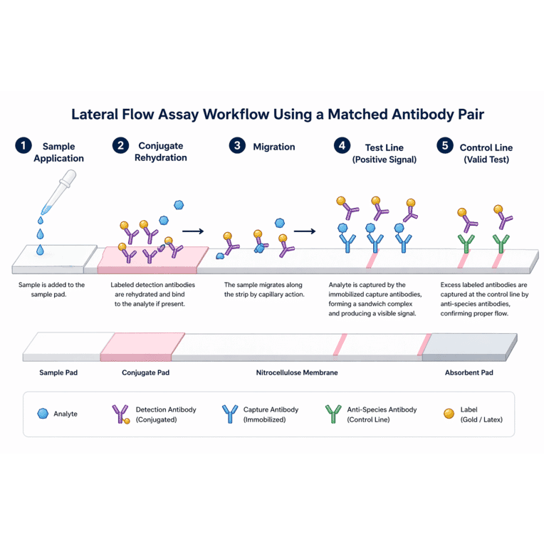
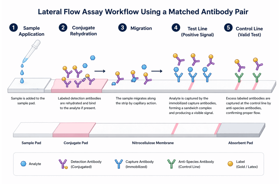
Schematic representation of a lateral flow assay using a matched antibody pair. Following application to the sample pad, the sample rehydrates labeled detection antibodies, which bind the target analyte if present. The resulting complexes migrate along the nitrocellulose membrane by capillary action. At the test line, immobilized capture antibodies bind the analyte-detection antibody complex, forming a sandwich and generating a visible signal. Excess labeled detection antibodies continue to migrate and are captured at the control line by anti-species antibodies, confirming proper assay function, while the absorbent pad maintains continuous flow across the strip. The analyte is represented by a blue shape, the detection antibody by a purple Y conjugated to a gold particle, the capture antibody by a blue Y, the control line antibody by a green Y, and the label by a gold particle (e.g. colloidal gold or latex).
Publishing research using Anti-Respiratory Syncytial Virus Matched Antibody Pair (A113172)? Please let us know so that we can list the citation on this page.