Human TSH ELISA Kit is a sandwich Enzyme-Linked Immunosorbent Assay (sELISA) designed for the in vitro quantitative determination of human TSH in serum, plasma, and cell culture supernatant.
Assay Type
Sandwich (quantitative)
Principle of Assay
Human TSH ELISA Kit (A334835) employs a one-step sandwich enzyme-linked immunosorbent assay (ELISA) for the quantitative determination of human TSH in serum, plasma, and cell culture supernatant. A 96-well microtiter plate is provided pre-coated with an antibody specific for TSH. Standards and test samples are added to the wells, allowing TSH to bind to the immobilized capture antibody. A prepared antibody mixture is added and incubated to form antibody–antigen complexes. After incubation, unbound material is removed by washing, and TMB substrate is added to develop colour through an HRP-catalysed reaction. The reaction is stopped with acidic stop solution, producing a yellow endpoint signal. The intensity of the colour is proportional to the amount of TSH captured in each well. The concentration of TSH is determined by measuring absorbance at 450 nm and interpolating from the standard curve.
Components
This ELISA Kit contains 10X 600 µL Capture Antibodies, 10X 600 µL Detector Antibodies, 2 Lyophilized Recombinant Protein Vials, 30 mL Assay Diluent #12Y, 10X 20 mL Wash Buffer, 12 mL TMB Solution, 12 mL Stop Solution, Pre-Coated 96-Well H-Microplate (12 × 8 well strips), and 2 Plate Seals.
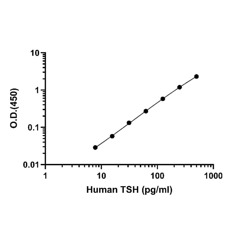
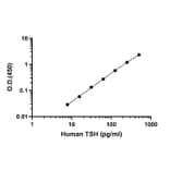
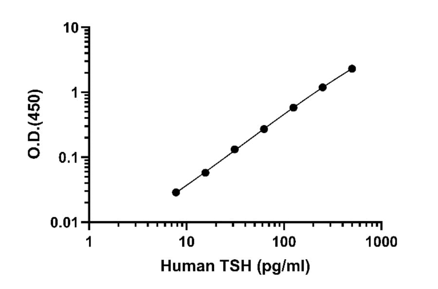
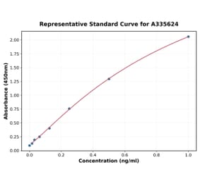
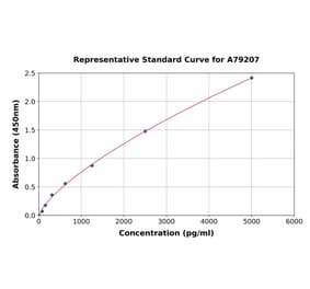
The standard curve for human thyroid-stimulating hormone (TSH) was generated by plotting optical density (O.D. 450 nm) versus TSH concentration (pg/mL) in Assay Diluent #12Y on a log-log scale. The curve demonstrates a strong linear relationship across the tested range, indicating reliable assay sensitivity and quantification accuracy for TSH detection.
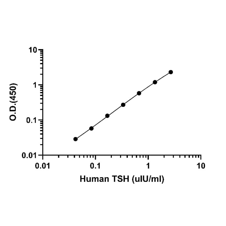
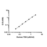
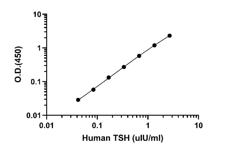
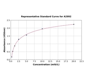
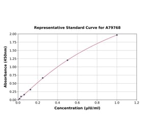
The standard curve for human thyroid-stimulating hormone (TSH) was established by plotting optical density (O.D. 450 nm) against TSH concentration (µIU/mL) in Assay Diluent #12Y on a log-log scale. The curve shows a strong linear correlation across the tested range, demonstrating the assay’s accuracy and sensitivity for quantifying TSH levels.
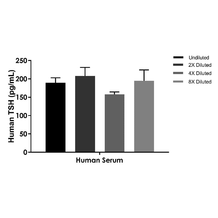
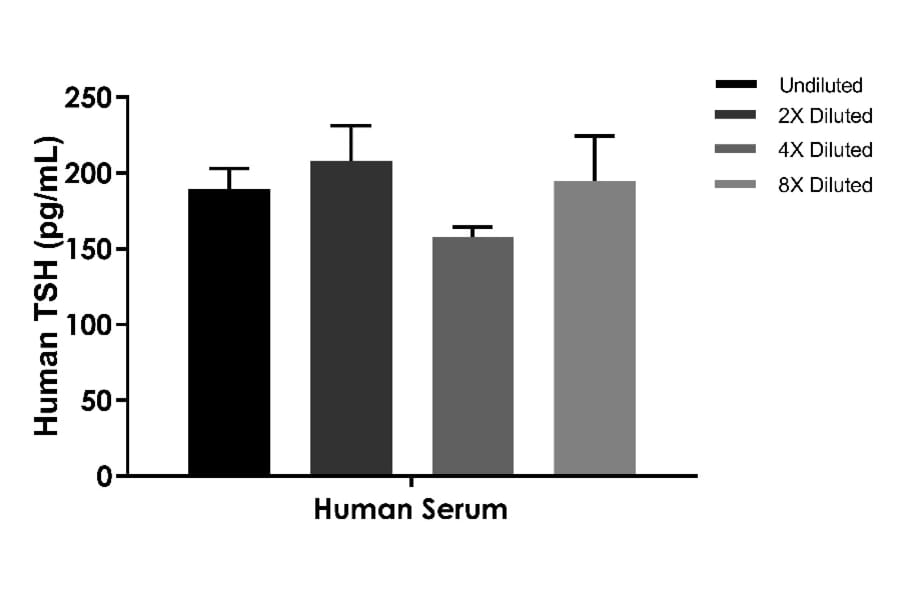
Human serum samples were analyzed for thyroid-stimulating hormone (TSH) concentrations using an ELISA assay at different dilution factors (undiluted, 2×, 4×, and 8×). The results show consistent detection of TSH across dilutions, with minimal variation, demonstrating the assay’s robustness and dilution linearity for serum samples. Data are expressed as mean ± SD.
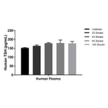
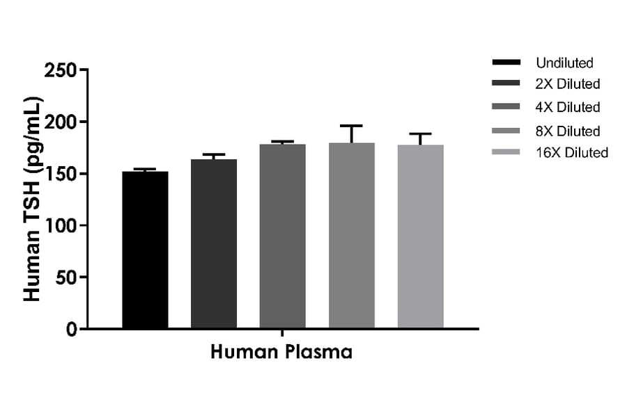
Human plasma samples were tested for thyroid-stimulating hormone (TSH) concentrations using an ELISA at different dilution factors (undiluted, 2×, 4×, 8×, and 16×). The results demonstrate consistent TSH detection across dilutions, indicating good assay performance and dilutional linearity in plasma matrices. Data are presented as mean ± SD.
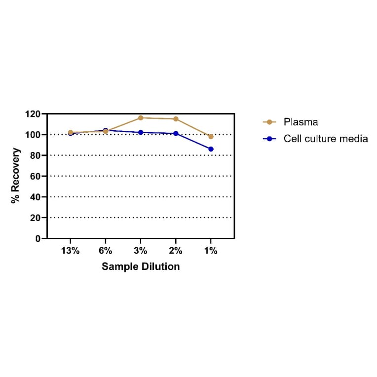
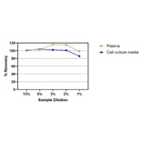
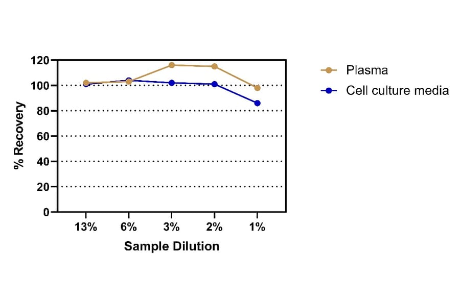
Percentage recovery of human TSH was assessed at various dilution levels (13%, 6%, 3%, 2%, and 1%) for plasma (brown line) and cell culture media (blue line). Both sample types showed acceptable recovery across dilutions, with plasma demonstrating slightly higher recovery consistency compared to cell culture media.
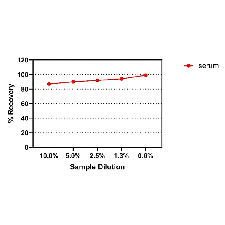
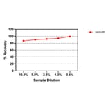
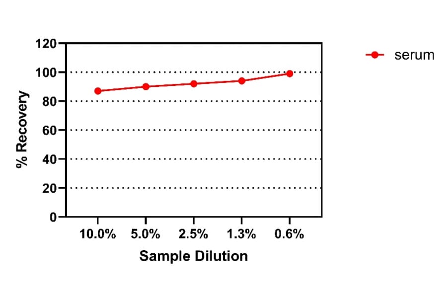
Percentage recovery of human TSH was evaluated at different dilution levels (10.0%, 5.0%, 2.5%, 1.3%, and 0.6%) in serum samples. Recovery values remained consistent across dilutions, indicating reliable assay performance and minimal matrix interference.
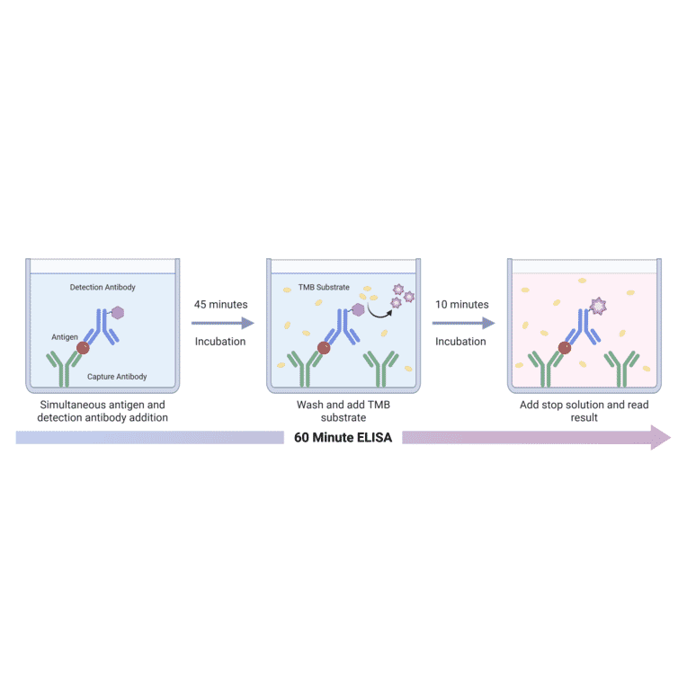
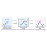
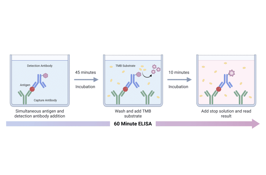
Schematic overview of the 60 Minute ELISA workflow illustrating antigen capture and colorimetric signal development. The target antigen is captured on the solid phase by an immobilized capture antibody while an HRP conjugated detection antibody binds simultaneously, forming a ternary immune complex during a 45 minute incubation. After washing to remove unbound components, tetramethylbenzidine (TMB) substrate is added. The horseradish peroxidase enzyme catalyzes oxidation of TMB, generating a blue reaction product that reflects the amount of bound antigen. Addition of stop solution terminates the reaction and converts the signal to a stable yellow color, which is measured spectrophotometrically after a 10 minute incubation. The entire assay is completed within approximately 60 minutes.