Immunofluorescence - Anti-14-3-3 eta Antibody [3G12] (A85361)
Immunofluorescent analysis of HeLa cells stained with Anti-14.3.3 eta Antibody, at a dilution of 1:1,000, in red. Blue is DAPI staining of nuclear DNA. The Anti-14.3.3 eta Antibody reveals the diffuse cytoplasmic distribution of 14.3.3? protein with higher concentration in the perinuclear region.
Western Blot - Anti-14-3-3 eta Antibody [3G12] (A85361)
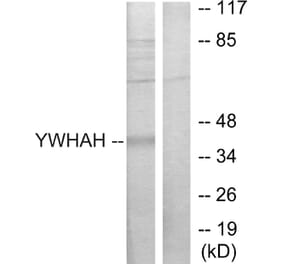
Western blot analysis of whole brain lysates (Lanes 2 & 3) and cell lysates (Lanes 4-8) using Anti-14.3.3 eta Antibody, at a dilution of 1:5,000, in green,: [Lane 1] protein standard (red), [Lane 2] rat brain, [Lane 3] mouse brain, [Lane 4] NIH-3T3, [Lane 5] HEK293, [Lane 6] HeLa, [Lane 7] SH-SY5Y, [Lane 8] C6 cells. Strong band at 28kDa corresponds to 14.3.3? protein, expressed in all preparations.
Immunohistochemistry - Anti-14-3-3 eta Antibody [3G12] (A85361)
Immunohistochemistry analysis of a 4% PFA fixed paraffin embedded rat hippocampus section with Anti-14-3-3 eta Antibody [3G12] (A85361) at a dilution of 1:1,000 detected with DAB (brown) using the Vector Labs ImmPRESS method and reagents with citra buffer retrieval. Counterstained with Hematoxylin (blue). Within the hippocampus, the Anti-14-3-3 eta Antibody [3G12] (A85361) labels the cytoplasm of neurons and their processes. Note: this antibody performs well in testing with both 4% PFA and standard NBF fixed tissues but does not stain long term NBF fixed tissue effectively.
Western Blot - Anti-14-3-3 eta Antibody [3G12] (A85361)
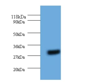
Blot of 20µg crude HeLa cell homogenate (lane 1) and 20µg rat brain lysate (lane2) was probed with Anti-14.3.3 eta Antibody, at a dilution of 1:2500). The Anti-14.3.3 eta Antibody recognizes 14.3.3? at about 28 kDa in both HeLa cell and rat brain lysates. 14.3.3? is more abundant in rat brain than in HeLa cells.
Publishing research using Anti-14-3-3 eta Antibody [3G12] (A85361)? Please let us know so that we can list the citation on this page.
Alternative products to Anti-14-3-3 eta Antibody [3G12] (A85361)


![Immunofluorescence - Anti-14-3-3 eta Antibody [3G12] (A85361) - Antibodies.com](https://cdn.antibodies.com/image/catalog/85/A85361_1.jpg?profile=product_top)
![Western Blot - Anti-14-3-3 eta Antibody [3G12] (A85361) - Antibodies.com](https://cdn.antibodies.com/image/catalog/85/A85361_2.jpg?profile=product_top)
![Immunohistochemistry - Anti-14-3-3 eta Antibody [3G12] (A85361) - Antibodies.com](https://cdn.antibodies.com/image/catalog/85/A85361_3.jpg?profile=product_top)
![Western Blot - Anti-14-3-3 eta Antibody [3G12] (A85361) - Antibodies.com](https://cdn.antibodies.com/image/catalog/85/A85361_5.jpg?profile=product_top)
![Immunofluorescence - Anti-14-3-3 eta Antibody [3G12] (A85361) - Antibodies.com](https://cdn.antibodies.com/image/catalog/85/A85361_1.jpg?profile=product_top_thumb)
![Western Blot - Anti-14-3-3 eta Antibody [3G12] (A85361) - Antibodies.com](https://cdn.antibodies.com/image/catalog/85/A85361_2.jpg?profile=product_top_thumb)
![Immunohistochemistry - Anti-14-3-3 eta Antibody [3G12] (A85361) - Antibodies.com](https://cdn.antibodies.com/image/catalog/85/A85361_3.jpg?profile=product_top_thumb)
![Western Blot - Anti-14-3-3 eta Antibody [3G12] (A85361) - Antibodies.com](https://cdn.antibodies.com/image/catalog/85/A85361_5.jpg?profile=product_top_thumb)
![Immunofluorescence - Anti-14-3-3 eta Antibody [3G12] (A85361) - Antibodies.com](https://cdn.antibodies.com/image/catalog/85/A85361_1.jpg?profile=product_image)
![Western Blot - Anti-14-3-3 eta Antibody [3G12] (A85361) - Antibodies.com](https://cdn.antibodies.com/image/catalog/85/A85361_2.jpg?profile=product_image)
![Immunohistochemistry - Anti-14-3-3 eta Antibody [3G12] (A85361) - Antibodies.com](https://cdn.antibodies.com/image/catalog/85/A85361_3.jpg?profile=product_image)
![Western Blot - Anti-14-3-3 eta Antibody [3G12] (A85361) - Antibodies.com](https://cdn.antibodies.com/image/catalog/85/A85361_5.jpg?profile=product_image)