Chicken polyclonal antibody to Adenylate Cyclase 3.
Applications
WB, ICC/IF
Dilutions
WB: 1:500-1:1,000, ICC/IF: 1:5,000-1:10,000
Reactivity
Rat, Mouse
Immunogen
C-terminal peptide of rat Adenylate Cyclase 3, with a Cys added to the N-terminus to allow coupling to KLH.
Sequence
PAAFPNGSSVTLPHQVVDNP
Host
Chicken
Clonality
Polyclonal
Isotype
IgY
Conjugate
Unconjugated
Purification
Immunogen affinity purification.
Concentration
1 mg/ml
Molecular Weight
~120 kDa (and above)
Product Form
Liquid
Formulation
Supplied in Phosphate Buffered Saline with 50% Glycerol and 5mM Sodium Azide.
Storage
Shipped at 4°C. Store at +4°C. Do not freeze!
Synonyms
AC-III, AC3, ADCY3, ADCY3_HUMAN, Adenylate cyclase, Adenylate cyclase type 3, Adenylate cyclase type III, Adenylyl cyclase 3, ATP pyrophosphate lyase, ATP pyrophosphate-lyase 3, olfactive type
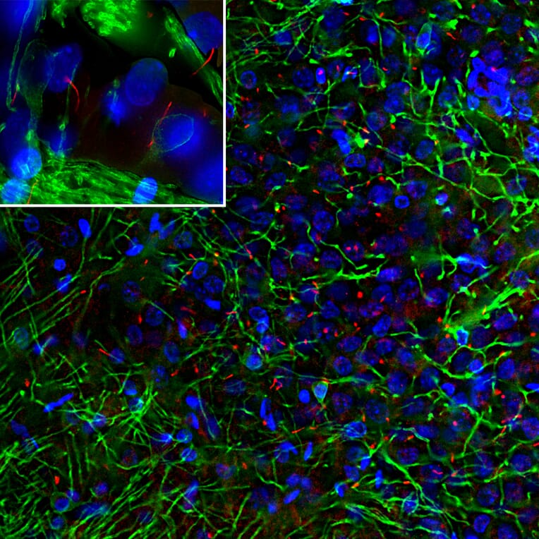
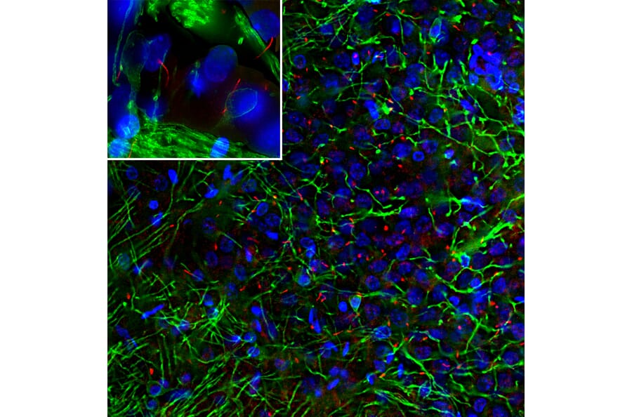
Immunofluorescent analysis of rat cortex section stained with Anti-Adenylate Cyclase 3 Antibody (A104341), at a dilution of 1:10,000, in red, and co-stained with Anti-CNPase Antibody [1H10] (A85413), at a dilution of 1:1,000, in green. The blue is Hoechst staining of nuclear DNA. Anti-Adenylate Cyclase 3 Antibody (A104341) reveals neuronal cilia while Anti-CNPase Antibody [1H10] (A85413) stains oligodendrocytes and the myelin sheath around axons.
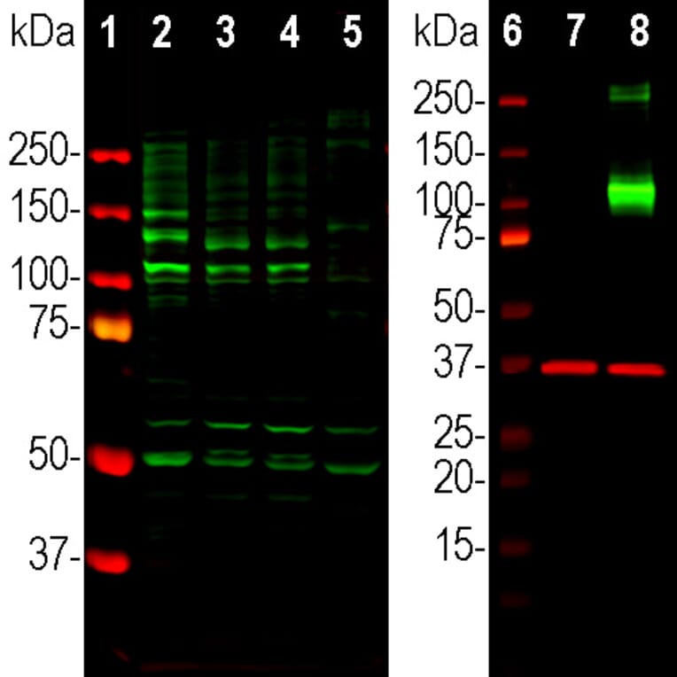
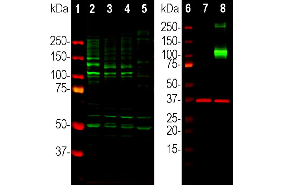
Western Blot - Anti-Adenylate Cyclase 3 Antibody (A104341)
Western blot analysis of different tissue lysates using Anti-Adenylate Cyclase 3 Antibody (A104341), at a dilution of 1:1,000, in green. Left: [Lane 1] protein standard, [Lane 2] rat hippocampus, [Lane 3] mouse hippocampus, [Lane 4] mouse frontal cortex, and [Lane 5] cow frontal cortex. Anti-Adenylate Cyclase 3 Antibody (A104341) detects variably glycosylated forms of ACIII protein with apparent molecular weights from ~120kDa and higher. Right: [Lane 6] protein standard, [Lane 7] non-transfected HEK293 cells, and [Lane 8] HEK293 cells transfected with DNA expressing Myc-DDK tagged full length human adenylate cyclase III from the appropriate cDNA (ACIII). The strong band at about 130kDa demonstrates overexpression of the ACIII protein, and those over 250kDa double band presumably corresponds to heavily glycosolated or aggregated forms of ACIII. The same blot was simultaneously probed with Anti-GAPDH Antibody [1D4] (A85382), at a dilution of 1:5,000, in red, which reveals the single GAPDH band at ~37kDa in both transfected and non-transfected cells.
Publishing research using Anti-Adenylate Cyclase 3 Antibody (A104341)? Please let us know so that we can list the citation on this page.