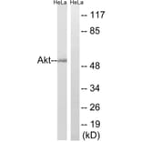
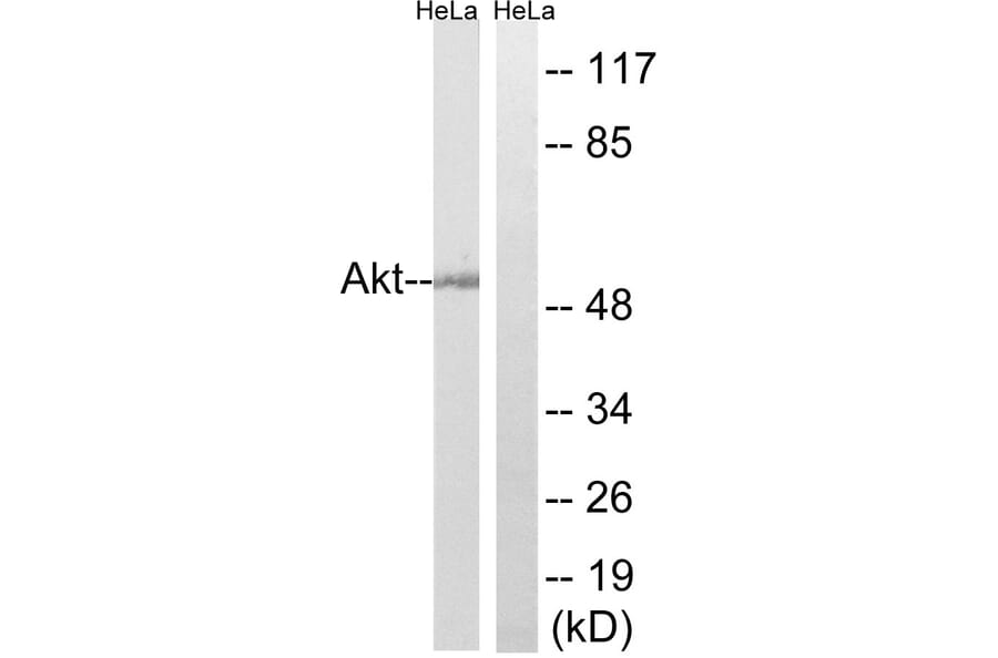
Western blot analysis of lysates from HeLa cells using Anti-Akt Antibody. The right hand lane represents a negative control, where the antibody is blocked by the immunising peptide.
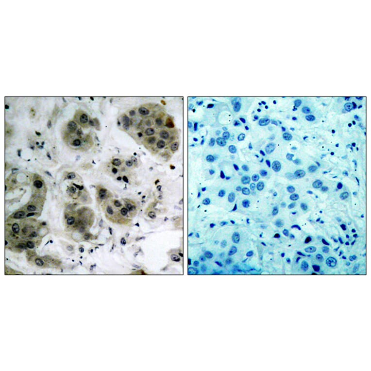
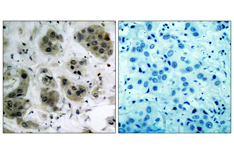
Immunohistochemical analysis of paraffin-embedded human breast carcinoma tissue using Anti-Akt Antibody. The right hand panel represents a negative control, where the antibody was pre-incubated with the immunising peptide.
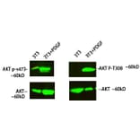
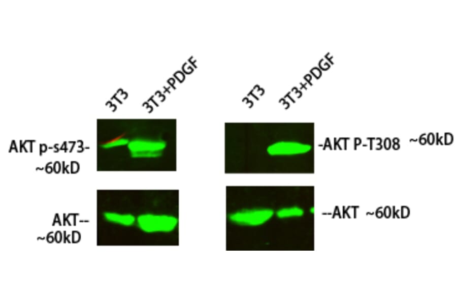
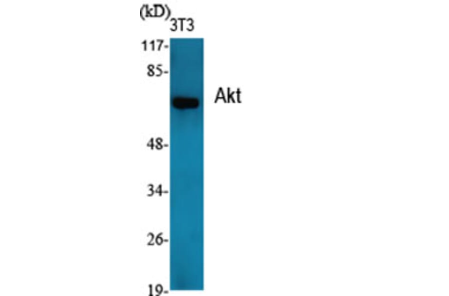
Western blot analysis of 3T3 cells treated with PDGF using Anti-Akt Antibody at 1:1,000 (4°C overnight). Goat Anti-Rabbit IgG (IRDye 800) was used as a secondary at 1:5,000 (25°C, 1 hour).