Recombinant construct corresponding to the C-terminal 398 amino acids of human Ankyrin 3 isotype 1 (NP_066267.2), expressed in and purified from E. coli. This segment is expressed by all three Ankyrin 3 isotypes and contains the DEATH domain sequence.
Host
Rabbit
Clonality
Polyclonal
Isotype
IgG
Conjugate
Unconjugated
Purification
Immunogen affinity purification.
Concentration
1 mg/ml
Molecular Weight
190 kDa, 270 kDa, 480 kDa
Product Form
Liquid
Formulation
Supplied in Phosphate Buffered Saline with 50% Glycerol and 5mM Sodium Azide.
Storage
Shipped at 4°C. Upon delivery aliquot and store at -20°C. Avoid freeze/thaw cycles.
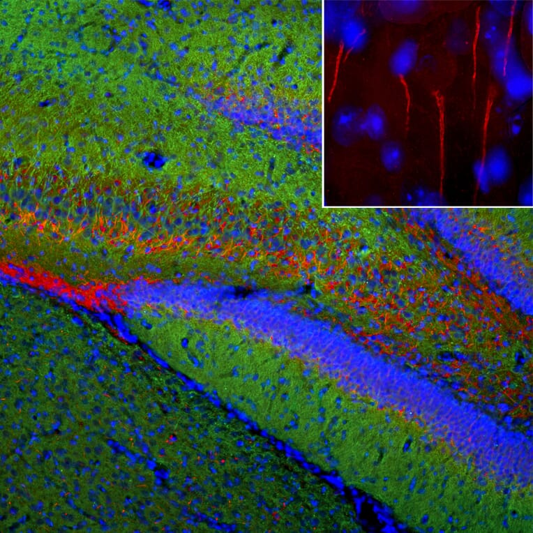
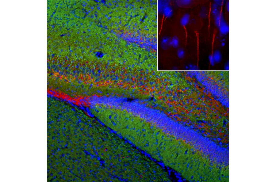
Immunofluorescent analysis of rat brain section stained with Anti-Ankyrin 3 Antibody (A104331), at a dilution of 1:1,000, in red, and co-stained with Anti-MAP2 Antibody [2C4] (A85459), at a dilution of 1:5,000, in green. The blue is Hoechst staining of nuclear DNA. Following transcardial perfusion of rat with 4% paraformaldehyde, brain was post fixed for 24 hours, cut to 45µM, and free-floating sections were stained with above antibodies. Anti-Ankyrin 3 Antibody (A104331) stains axonal initial segments, while Anti-MAP2 Antibody [2C4] (A85459) labels MAP2 protein expressed in the perikarya and dendrites of most neurons.
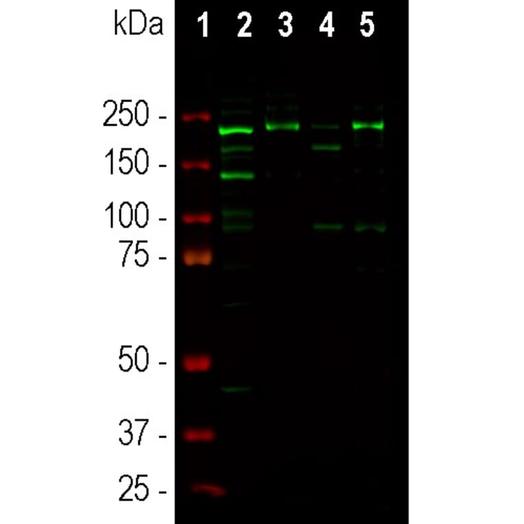
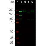
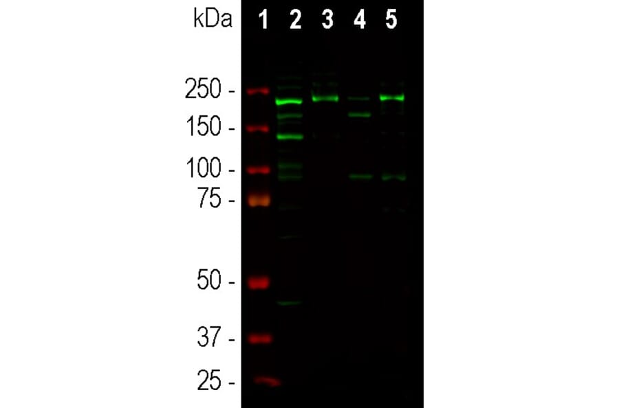
Western blot analysis of different tissue lysates using Anti-Ankyrin 3 Antibody (A104331), at a dilution of 1:1,000, in green. The lanes contain: [Lane 1] protein standard (red), [Lane 2] rat cortex, [Lane 3] rat cortex membrane enriched fraction, [Lane 4] mouse cortex, and [Lane 5] mouse cortex membrane enriched fraction. The band at ~190kDa corresponds to one of the three high molecular weight forms of Ankyrin 3; the 270kDa and 480kDa isoforms can be seen on longer exposure of the blot.
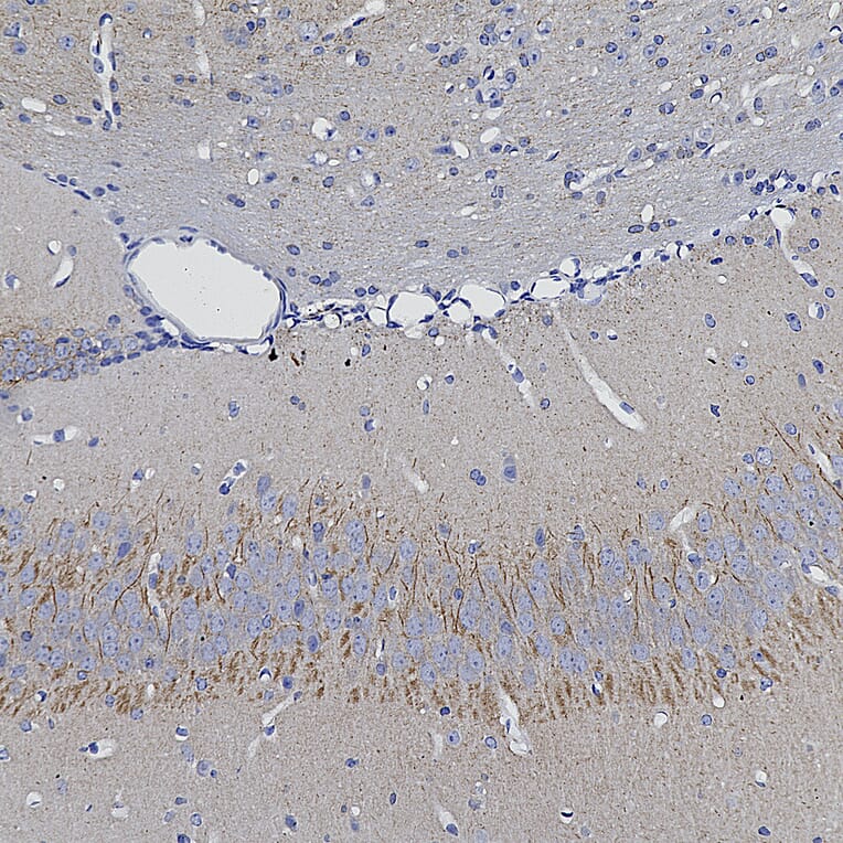
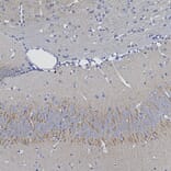
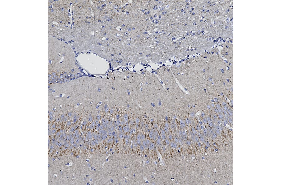
Immunohistochemistry analysis of a 4% PFA fixed paraffin embedded mouse hippocampus section with Anti-Ankyrin 3 Antibody (A104331) at a dilution of 1:2,000 detected with DAB (brown) using the Vector Labs ImmPRESS method and reagents with citra buffer retrieval. Counterstained with Hematoxylin (blue). Anti-Ankyrin 3 Antibody (A104331) labels axonal initial segments, the site of action potential initiation. Note: this antibody performs well in testing with both 4% PFA and standard NBF fixed mouse, human, and rat tissues.
Publishing research using Anti-Ankyrin 3 Antibody (A104331)? Please let us know so that we can list the citation on this page.