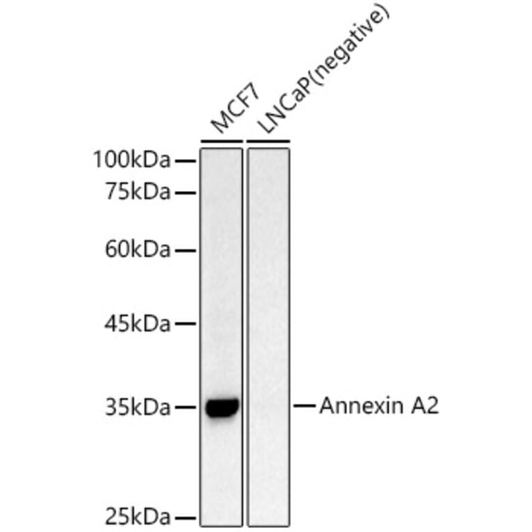
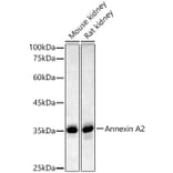
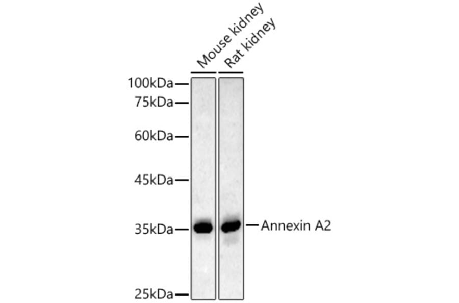
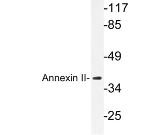
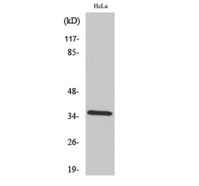
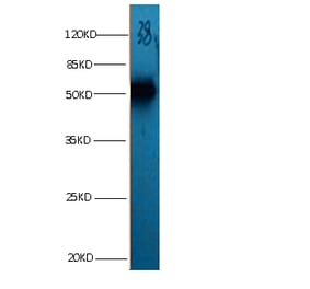
Annexin A2 expression in various lysates analyzed by western blot. Primary antibody incubation was performed for 1 hour on 25ug protein per lane with Anti-Annexin A2 Antibody (A13514) at a dilution of 1:500 and detected with chemiluminescence.
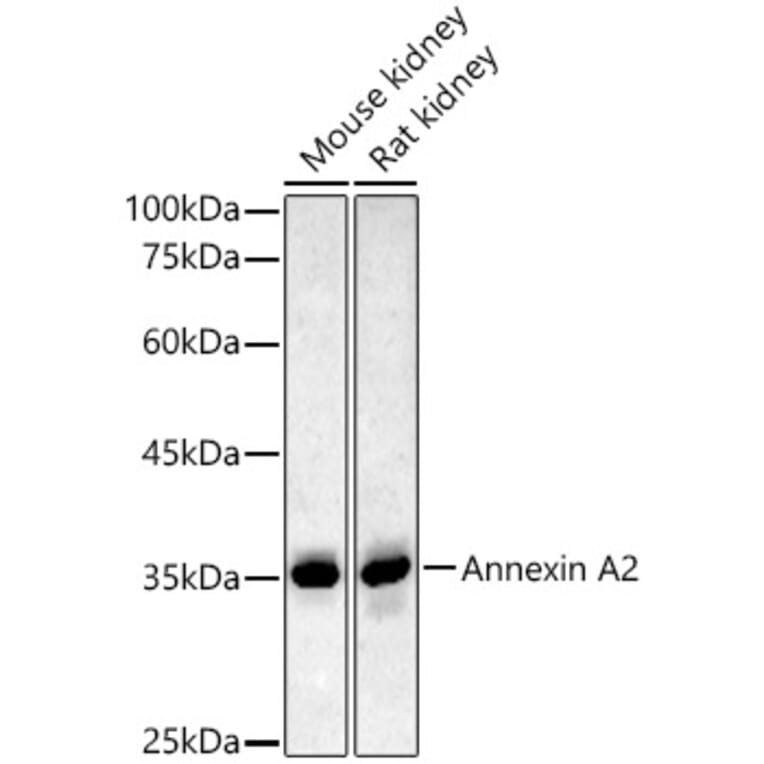
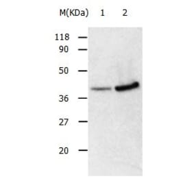
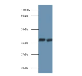
Annexin A2 expression in various lysates analyzed by western blot. Primary antibody incubation was performed for 1 hour on 25ug protein per lane with Anti-Annexin A2 Antibody (A13514) at a dilution of 1:500 and detected with chemiluminescence.
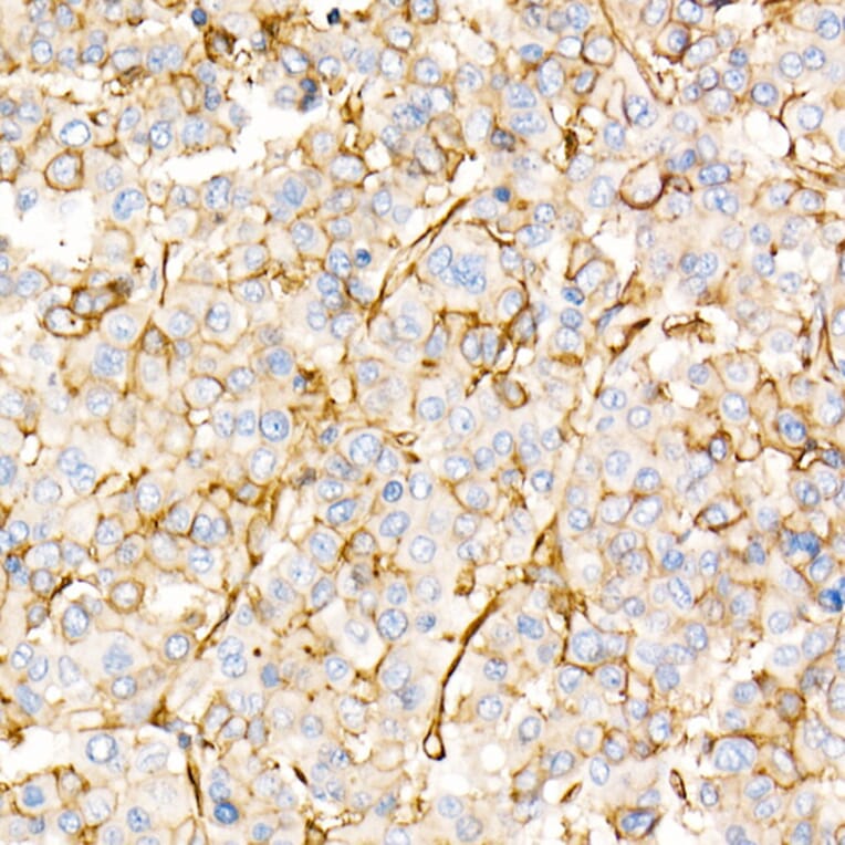
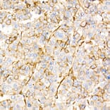
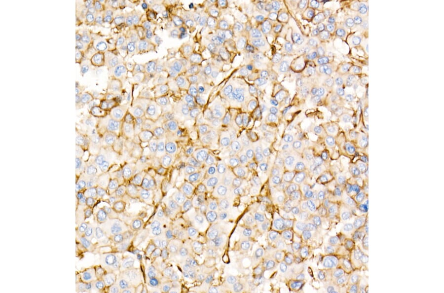
Annexin A2 expression in human liver cancer tissue analyzed by immunohistochemistry. Tissue was paraffin-embedded, and antigen retrieval was achieved with 10 mM citrate buffer, pH 6.0, under high pressure. Staining was performed with Anti-Annexin A2 Antibody (A13514) at a dilution of 1:20.
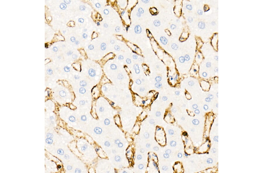
Annexin A2 expression in human liver tissue analyzed by immunohistochemistry. Tissue was paraffin-embedded, and antigen retrieval was achieved with 10 mM citrate buffer, pH 6.0, under high pressure. Staining was performed with Anti-Annexin A2 Antibody (A13514) at a dilution of 1:20.
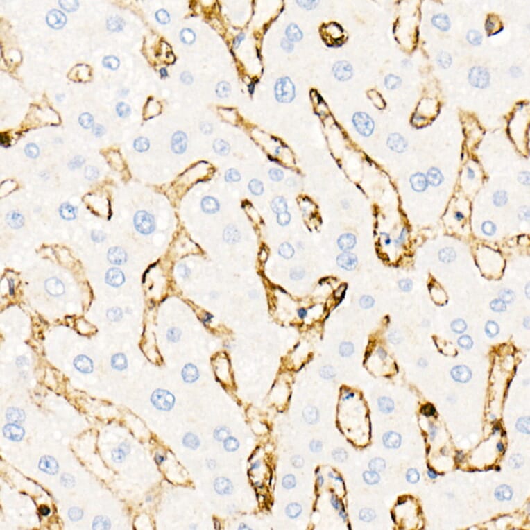
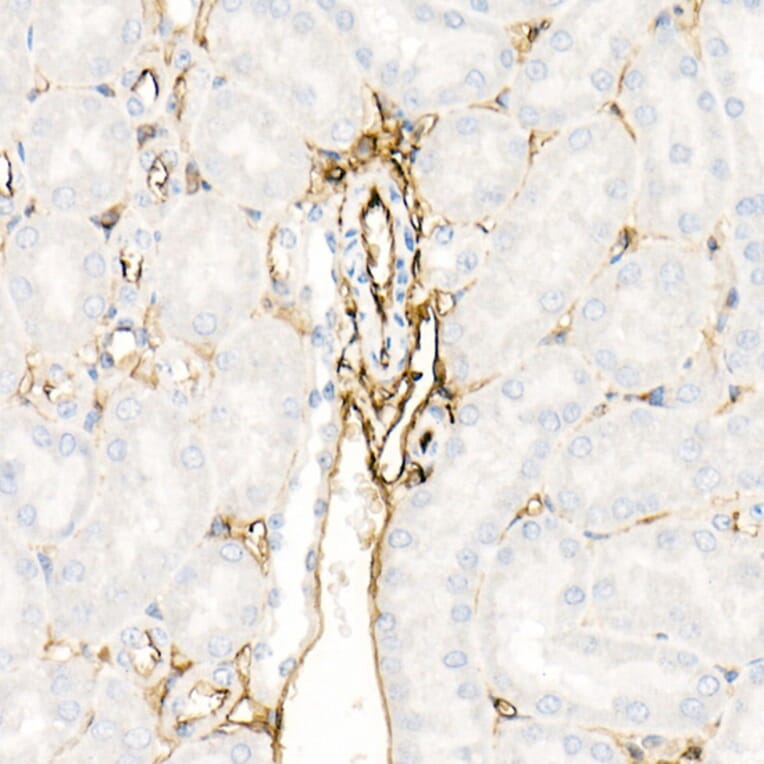
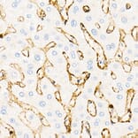
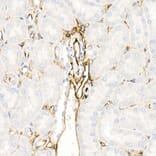
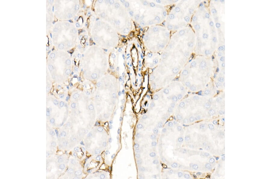
Annexin A2 expression in mouse kidney tissue analyzed by immunohistochemistry. Tissue was paraffin-embedded, and antigen retrieval was achieved with 10 mM citrate buffer, pH 6.0, under high pressure. Staining was performed with Anti-Annexin A2 Antibody (A13514) at a dilution of 1:20.
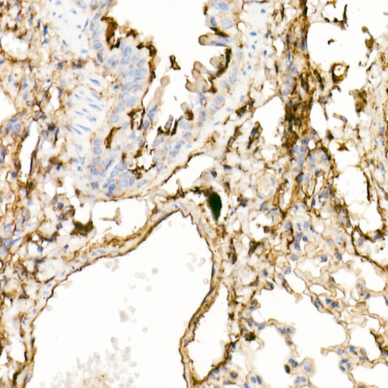
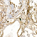
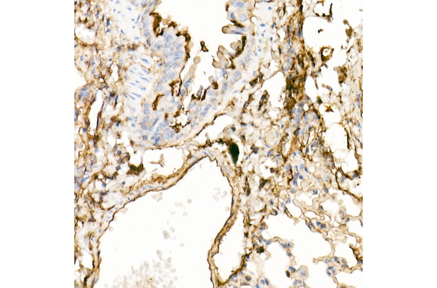
Annexin A2 expression in mouse lung tissue analyzed by immunohistochemistry. Tissue was paraffin-embedded, and antigen retrieval was achieved with 10 mM citrate buffer, pH 6.0, under high pressure. Staining was performed with Anti-Annexin A2 Antibody (A13514) at a dilution of 1:20.
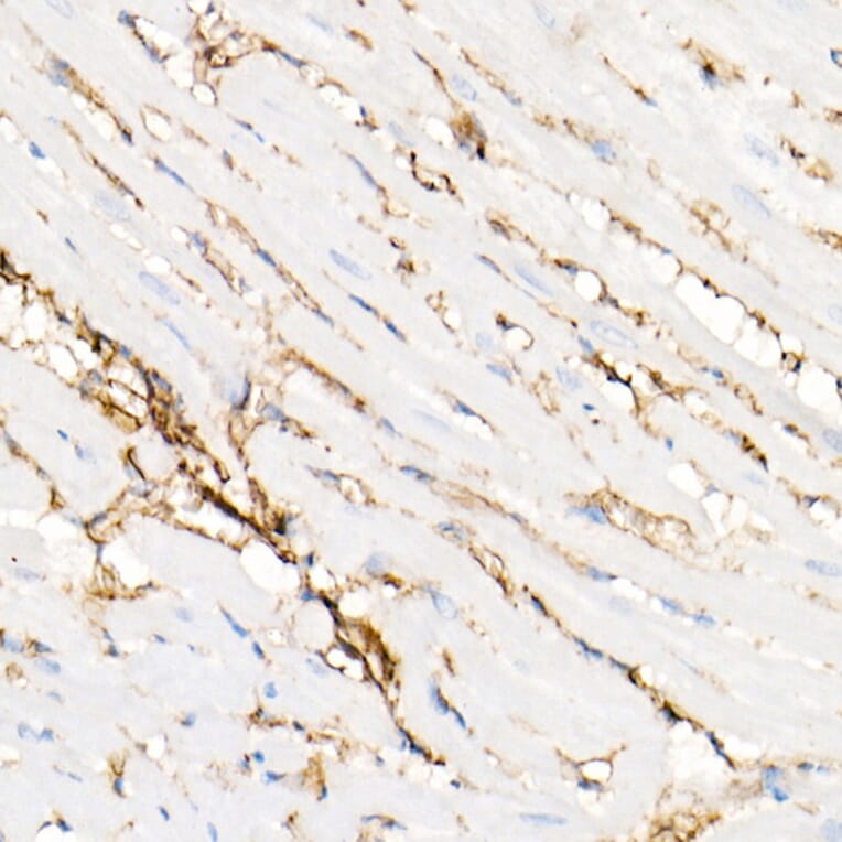
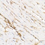
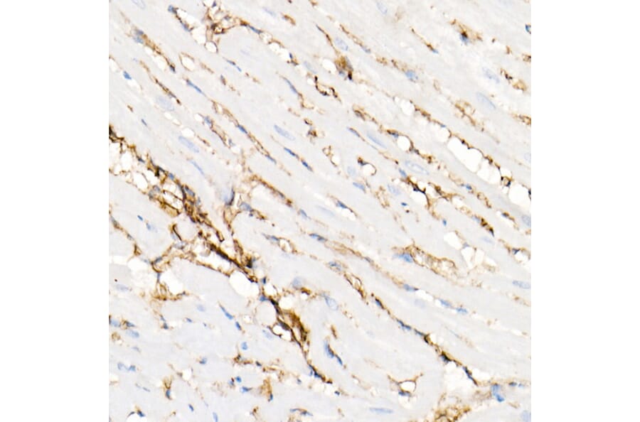
Annexin A2 expression in rat heart tissue analyzed by immunohistochemistry. Tissue was paraffin-embedded, and antigen retrieval was achieved with 10 mM citrate buffer, pH 6.0, under high pressure. Staining was performed with Anti-Annexin A2 Antibody (A13514) at a dilution of 1:20.
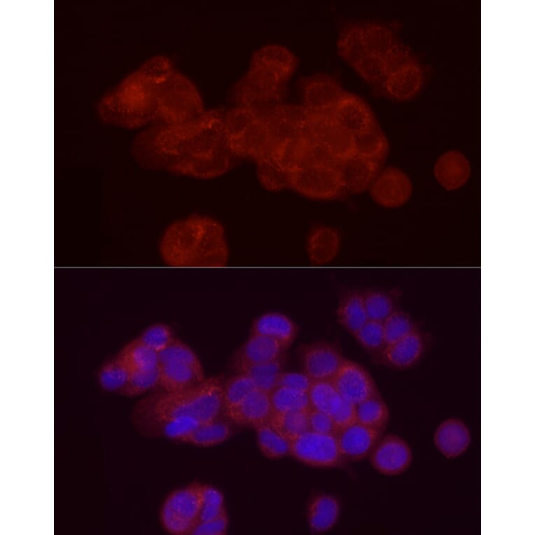
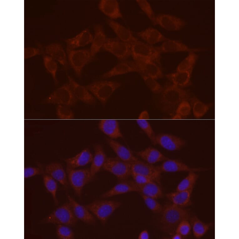
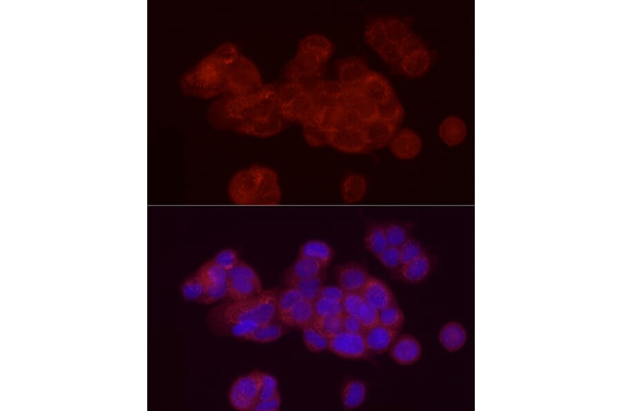
Annexin A2 expression in MCF7 cells analyzed by immunofluorescence. Staining was performed with Anti-Annexin A2 Antibody (A13514) at a dilution of 1:20 followed by Cy3-conjugated Goat anti-Rabbit IgG (H+L) secondary antibody at a dilution of 1:500. Nuclei were stained with DAPI (blue).
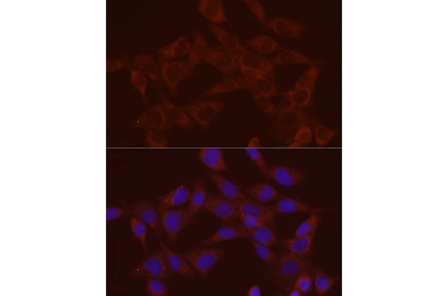
Annexin A2 expression in NIH/3T3 cells analyzed by immunofluorescence. Staining was performed with Anti-Annexin A2 Antibody (A13514) at a dilution of 1:20 followed by Cy3-conjugated Goat anti-Rabbit IgG (H+L) secondary antibody at a dilution of 1:500. Nuclei were stained with DAPI (blue).