Supplied in Phosphate Buffered Saline, pH 7.3, with 50% Glycerol and 0.02% Sodium Azide.
Storage
Shipped at 4°C. Upon delivery aliquot and store at -20°C. Avoid freeze/thaw cycles.
Synonyms
ARID domain-containing protein 3A, AT-rich interactive domain-containing protein 3A, B-cell regulator of IgH transcription, Bright, Dead ringer-like protein 1, DRIL1, DRIL3, DRX, E2F-binding protein 1, E2FBP1
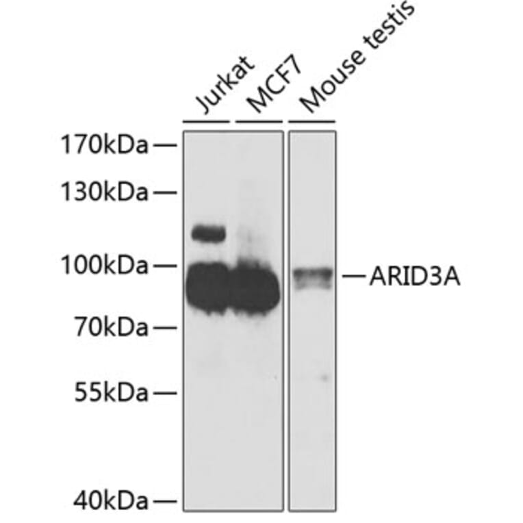
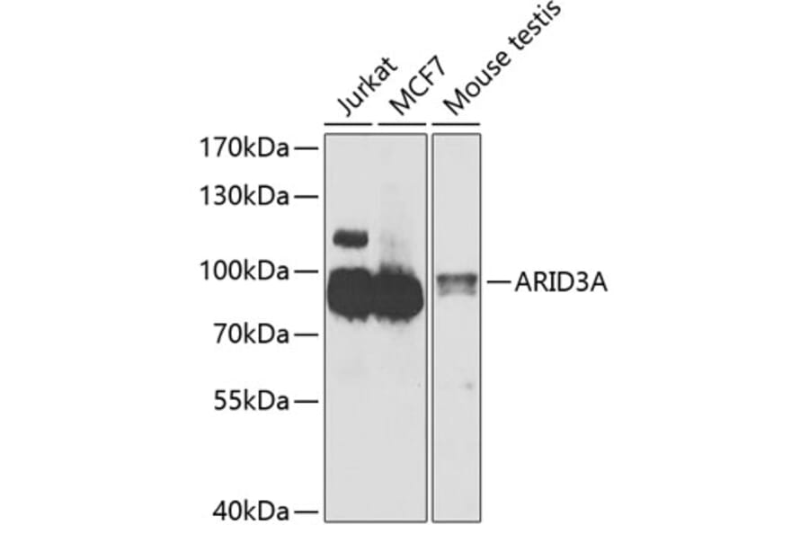
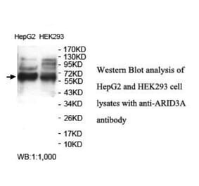
Western blot analysis of extracts of various cell lines, using Anti-ARID3A Antibody (A15831) at 1:1,000 dilution. The secondary antibody was Goat Anti-Rabbit IgG H&L Antibody (HRP) at 1:10,000 dilution. Lysates/proteins were present at 25µg per lane. The blocking buffer used was 3% non-fat dry milk in TBST. Detection was with a ECL Basic Kit. Exposure time: 90s.
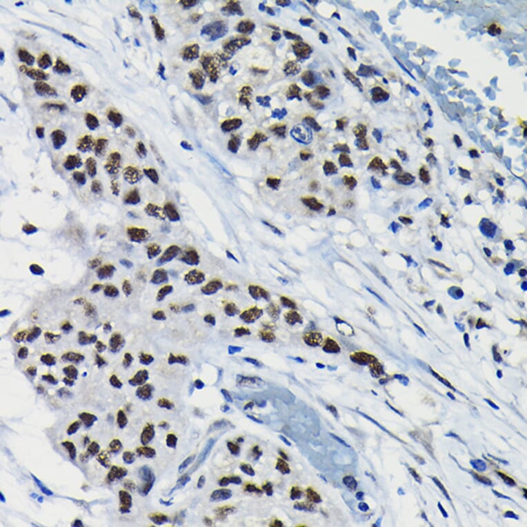
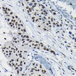
Immunohistochemistry analysis of paraffin-embedded rat lung using Anti-ARID3A Antibody (A15831) at a dilution of 1:100 (40x lens). Perform high pressure antigen retrieval with 10 mM citrate buffer pH 6.0 before commencing with IHC staining protocol.
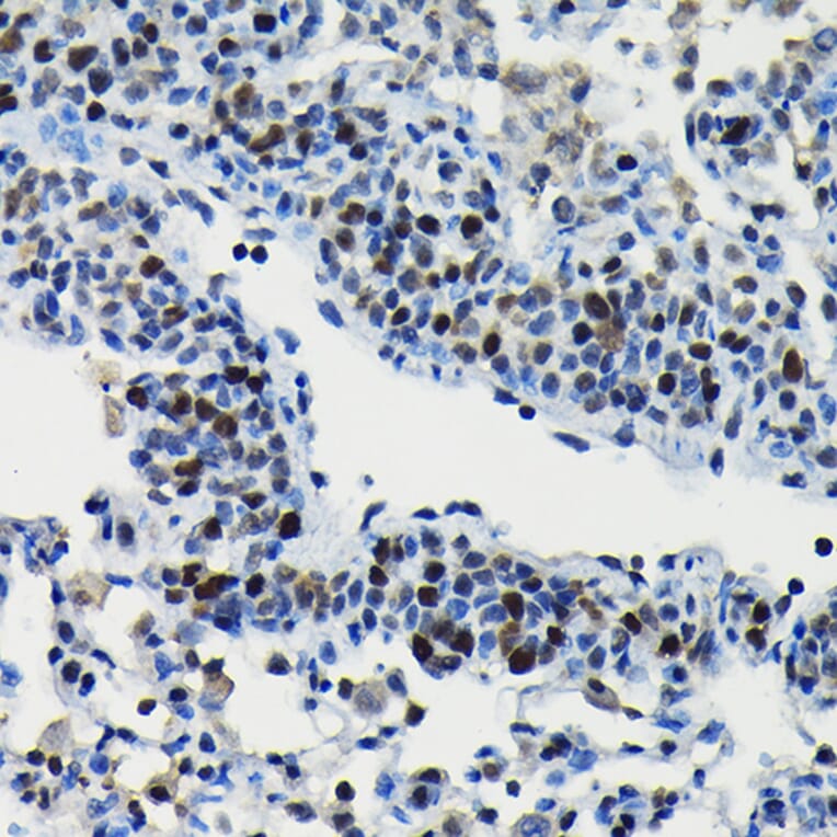
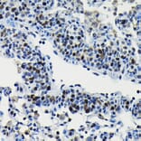
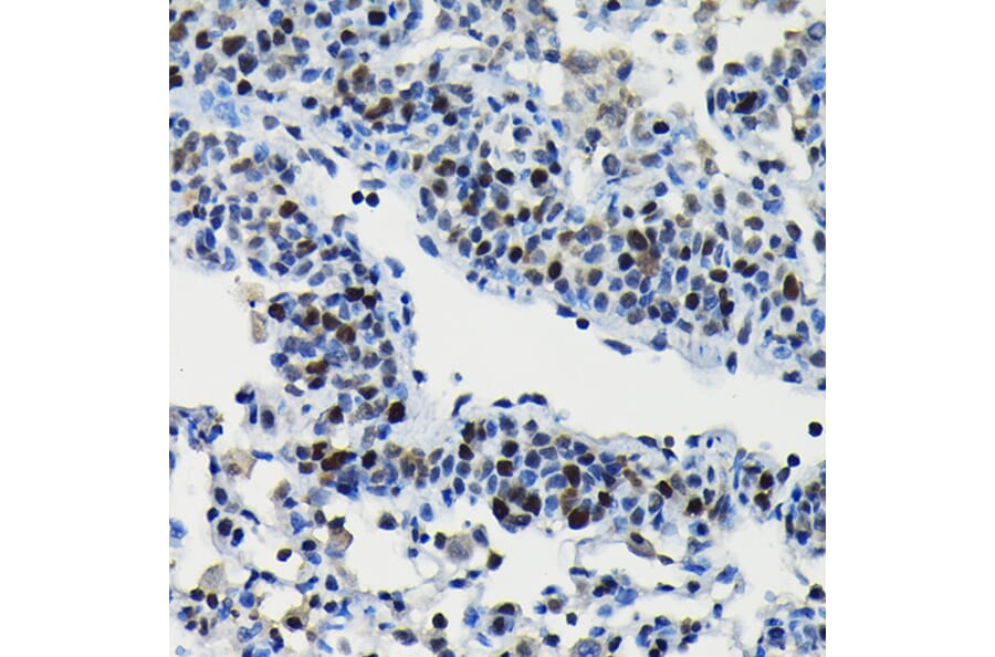
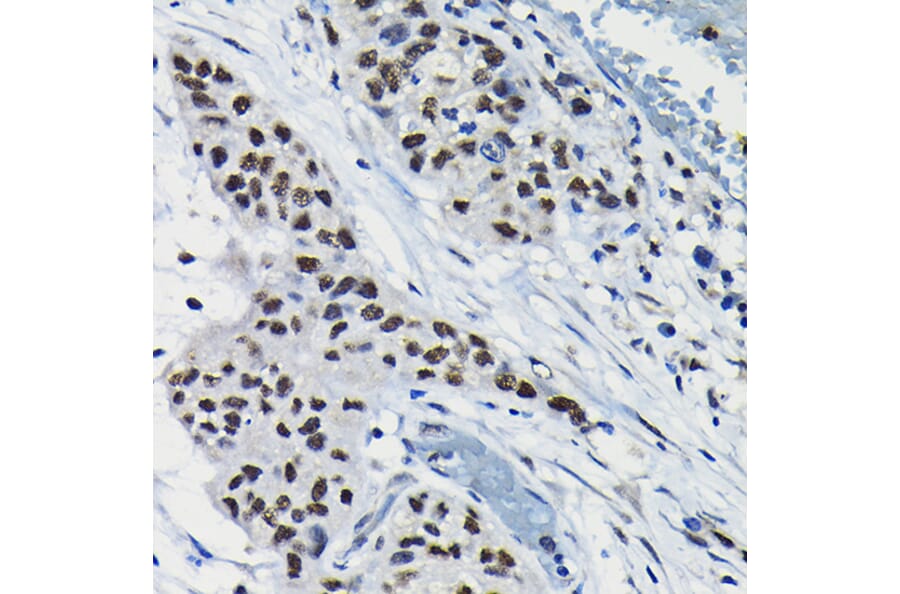
Immunohistochemistry analysis of paraffin-embedded human lung cancer using Anti-ARID3A Antibody (A15831) at a dilution of 1:100 (40x lens). Perform high pressure antigen retrieval with 10 mM citrate buffer pH 6.0 before commencing with IHC staining protocol.
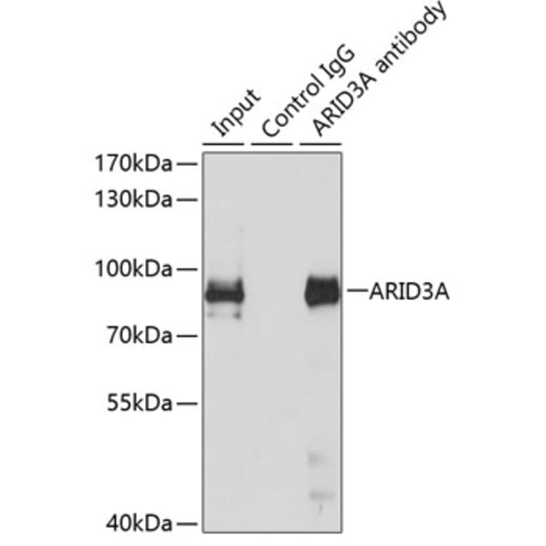
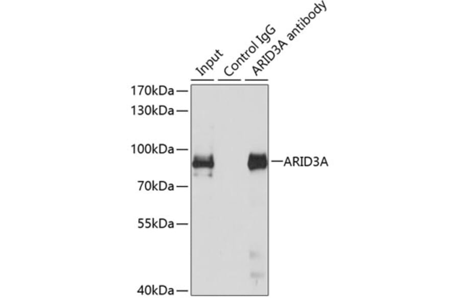
Immunoprecipitation analysis of 150µg extracts of MCF7 cells using 3µg of Anti-ARID3A Antibody (A15831). This Western blot was performed on the immunoprecipitate using Anti-ARID3A Antibody (A15831) at a dilution of 1:500.
Publishing research using Anti-ARID3A Antibody (A15831)? Please let us know so that we can list the citation on this page.
Alternative products to Anti-ARID3A Antibody (A15831)