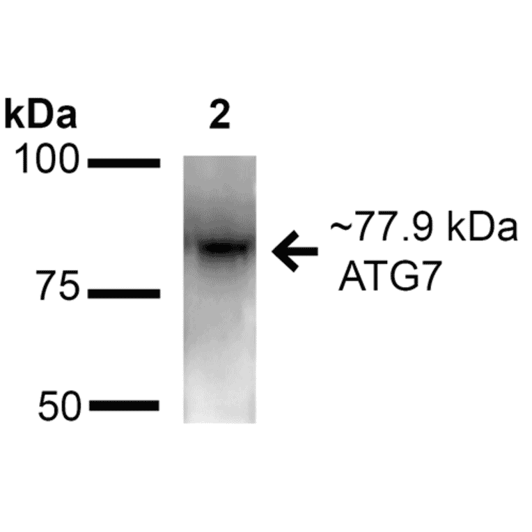
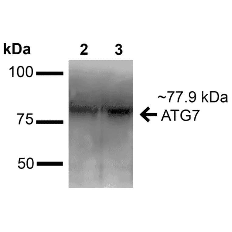
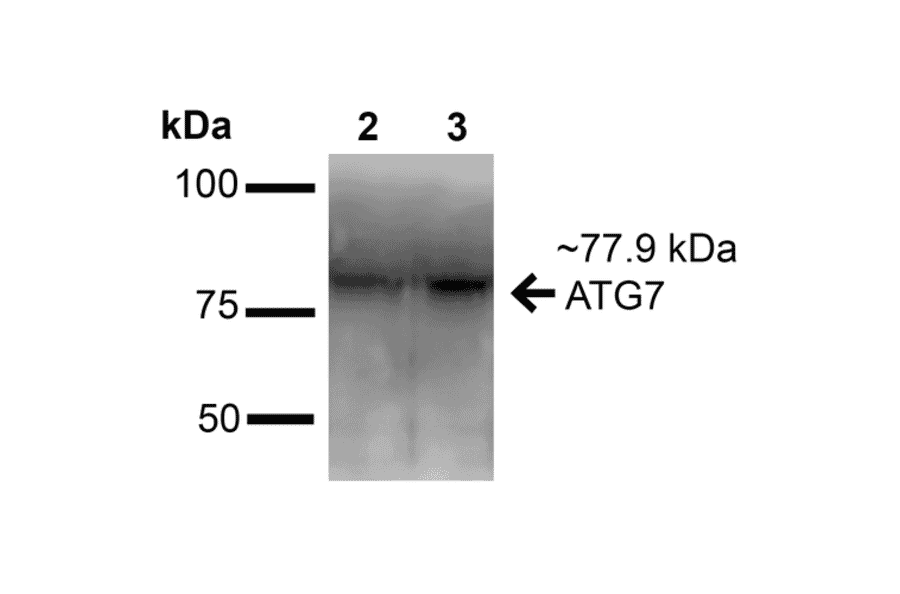
Western blot analysis of mouse and rat brain cell lysates showing detection of ~77.9 kDa ATG7 protein using Anti-ATG7 Antibody (A305057) at 1:1,000 for 16 hours at 4°C. Lane 1: Molecular Weight Ladder. Lane 2: mouse brain cell lysates. Lane 3: rat brain cell lysates. Load: 20 µg. Block: 2% BSA and 2% Skim Milk in 1X TBST. The secondary antibody used was Goat Anti-Rabbit IgG: HRP at 1:2000 for 60 minutes at room temperature. Color Development: ECL solution for 6 min at room temperature. Predicted/Observed Size: ~77.9 kDa.
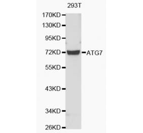
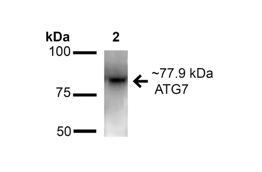
Western blot analysis of human Embryonic kidney epithelial cell line (HEK293T) lysate showing detection of ~77.9 kDa ATG7 protein using Anti-ATG7 Antibody (A305057) at 1:1,000 for 16 hours at 4°C. Lane 1: Molecular Weight Ladder (MW). Lane 2: human Embryonic kidney epithelial cell line (HEK293T) lysate. Load: 20 µg. Block: 2% BSA and 2% Skim Milk in 1X TBST. The secondary antibody used was Goat Anti-Rabbit IgG: HRP at 1:2000 for 60 minutes at room temperature. Color Development: ECL solution for 6 min at room temperature. Predicted/Observed Size: ~77.9 kDa.
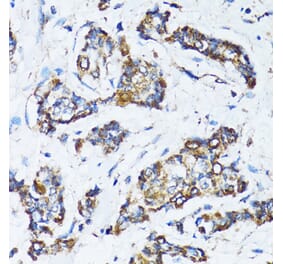
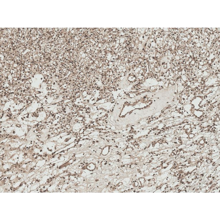
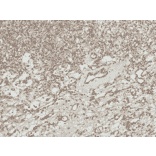
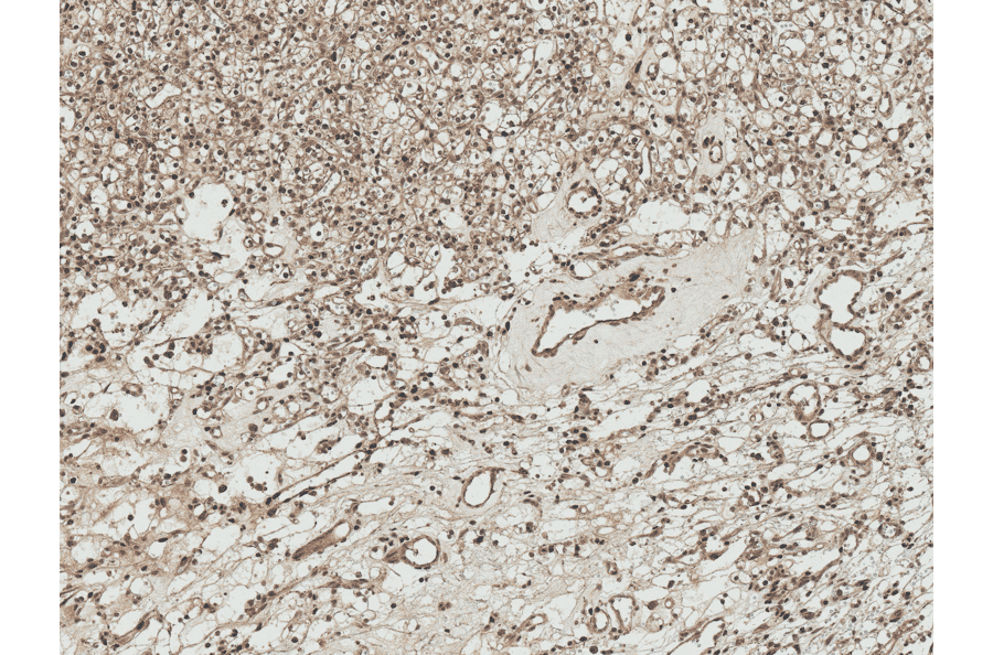
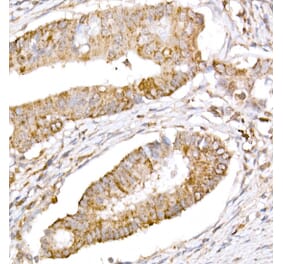
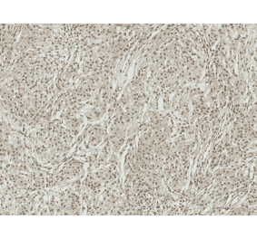
Immunohistochemistry analysis of human kidney, fixed in formalin and paraffin-embedded. The Primary Antibody used was Anti-ATG7 Antibody (A305057) at 1:50 for 30 minutes at room temperature. Counterstain: Hematoxylin. Magnification: 10X.
Publishing research using Anti-ATG7 Antibody (A305057)? Please let us know so that we can list the citation on this page.
Alternative products to Anti-ATG7 Antibody (A305057)


![Western Blot - Anti-ATG7 Antibody [ARC0083] (A306787) - Antibodies.com](https://cdn.antibodies.com/image/catalog/306/A306787_1.jpg?profile=product_alternative)