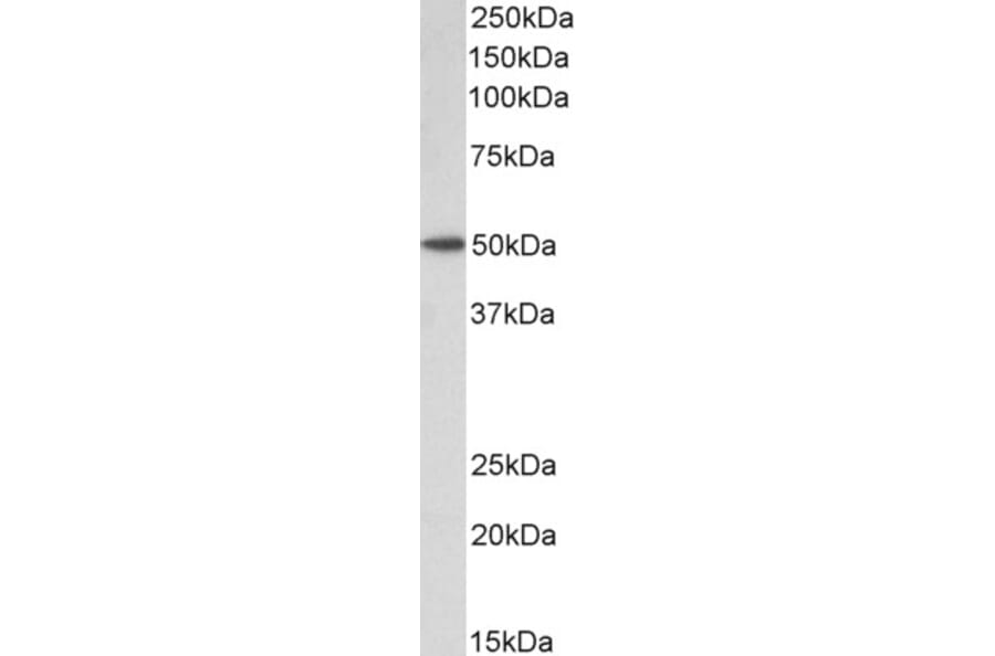
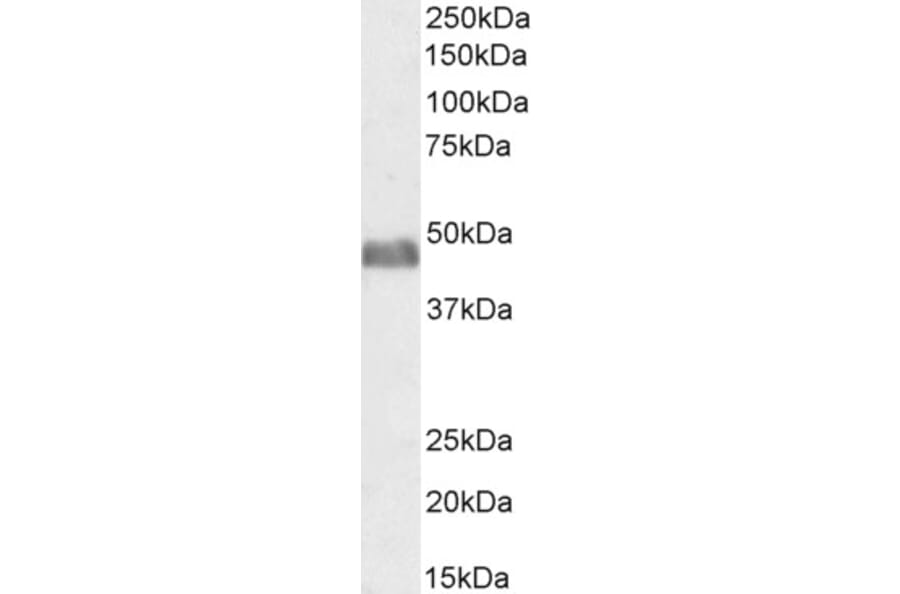
Western Blot - Anti-BAF53A + BAF53B Antibody (A83400)
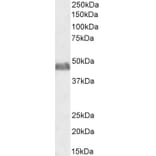
BAF53A + BAF53B expression in Rat Skeletal Muscle lysate analyzed by western blot. Cells were lysed in RIPA buffer and 35µg protein was run per lane. Primary antibody incubation was performed with Anti-BAF53A + BAF53B Antibody (A83400) at 2µg/ml and detected by chemiluminescence.
Western Blot - Anti-BAF53A + BAF53B Antibody (A83400)
BAF53A + BAF53B expression in NIH3T3 nuclear cell lysate analyzed by western blot. Cells were lysed in RIPA buffer and 35µg protein was run per lane. Primary antibody incubation was performed with Anti-BAF53A + BAF53B Antibody (A83400) at 0.5µg/ml and detected by chemiluminescence.
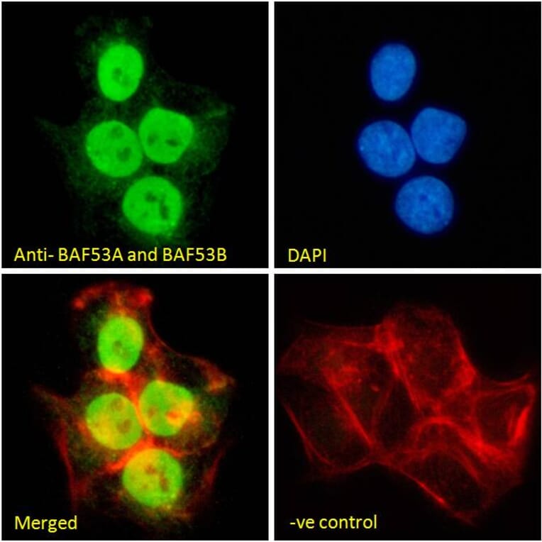
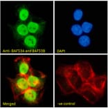
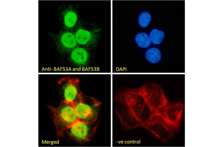
BAF53A + BAF53B expression in A431 cells analyzed by immunofluorescence. Cells were permeabilized with 0.15% Triton. Staining was performed with Anti-BAF53A + BAF53B Antibody (A83400) at 10µg/ml for 1 hour and Alexa Fluor 488 secondary antibody at 2µg/ml. Nuclear staining shown and nuclei were stained with DAPI (blue) while actin filaments were stained with phalloidin (red). Negative control: Goat IgG Isotype Control at 10µg/ml followed by Alexa Fluor 488 secondary antibody at 2µg/ml.
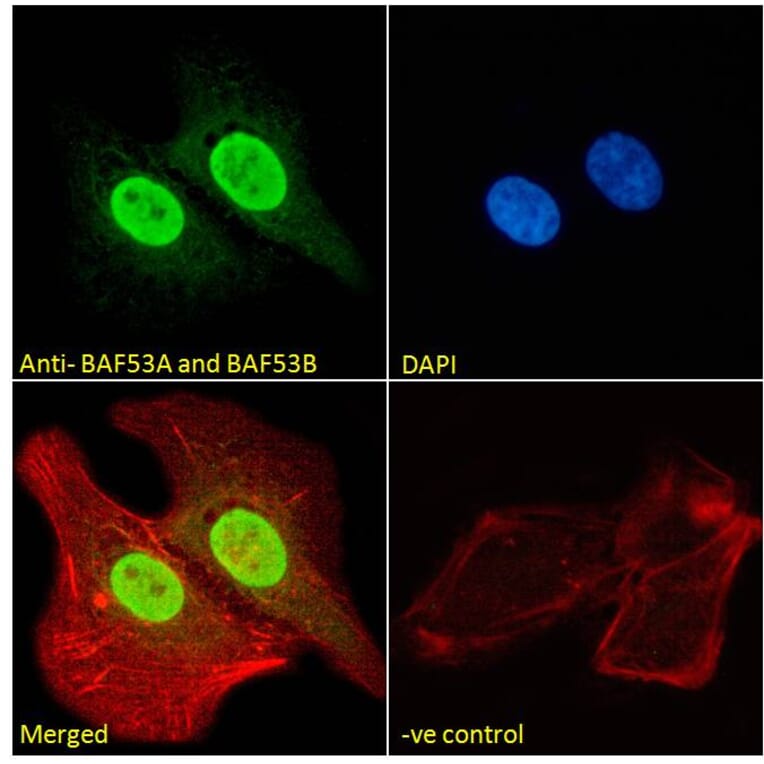
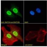
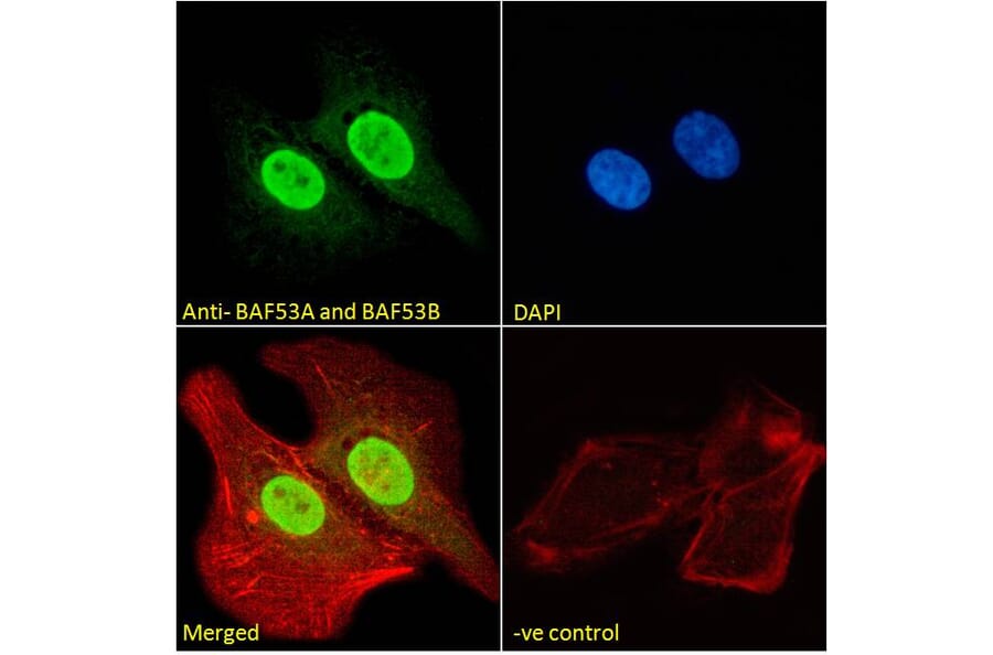
BAF53A + BAF53B expression in U2OS cells analyzed by immunofluorescence. Cells were permeabilized with 0.15% Triton. Staining was performed with Anti-BAF53A + BAF53B Antibody (A83400) at 10µg/ml for 1 hour and Alexa Fluor 488 secondary antibody at 2µg/ml. Nuclear staining shown and nuclei were stained with DAPI (blue) while actin filaments were stained with phalloidin (red). Negative control: Goat IgG Isotype Control at 10µg/ml followed by Alexa Fluor 488 secondary antibody at 2µg/ml.
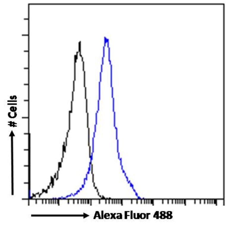
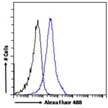
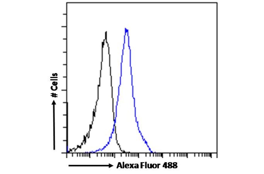
BAF53A + BAF53B expression in A431 cells (blue line) analyzed by flow cytometry. Cells were fixed in PFA and permeabilized with 0.5% Triton. Staining was performed with Anti-BAF53A + BAF53B Antibody (A83400) at 10µg/ml for 1 hour and Alexa Fluor 488 secondary antibody at 1µg/ml. Negative Control: Goat IgG Isotype Control (black line) followed by Alexa Fluor 488 secondary antibody.