Supplied in Phosphate Buffered Saline, pH 7.3, with 50% Glycerol and 0.02% Sodium Azide.
Storage
Shipped at 4°C. Upon delivery aliquot and store at -20°C. Avoid freeze/thaw cycles.
Synonyms
Breast carcinoma-amplified sequence 2, DAM1, DNA amplified in mammary carcinoma 1 protein, Pre-mRNA-splicing factor SPF27, Spliceosome-associated protein SPF 27
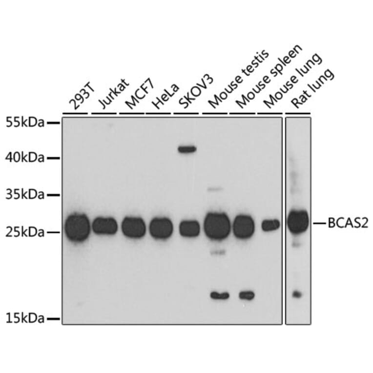
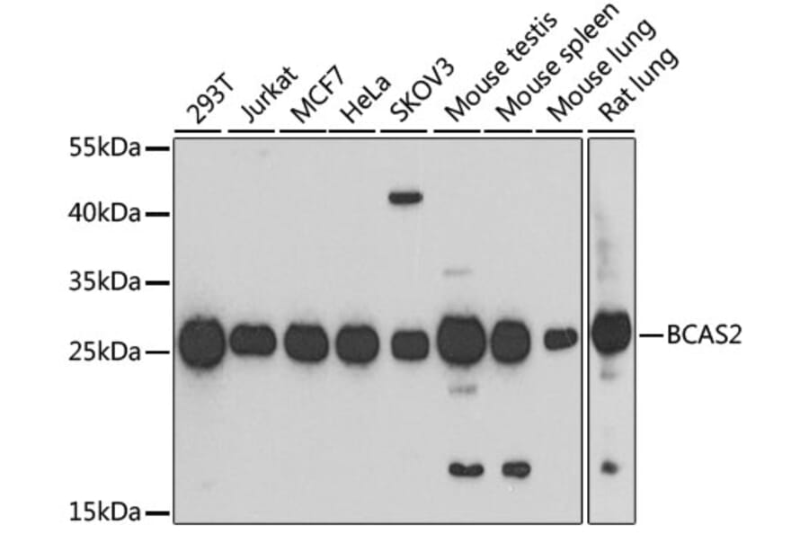
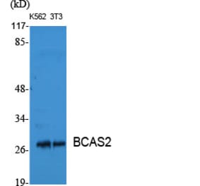
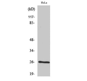
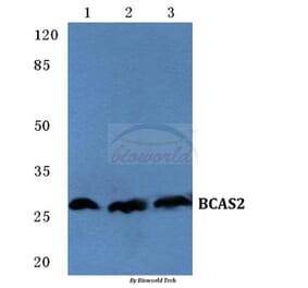
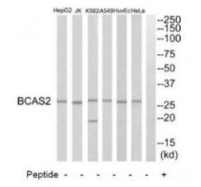
Western blot analysis of extracts of various cell lines, using Anti-BCAS2 Antibody (A14558) at 1:1,000 dilution. The secondary antibody was Goat Anti-Rabbit IgG H&L Antibody (HRP) at 1:10,000 dilution. Lysates/proteins were present at 25µg per lane. The blocking buffer used was 3% non-fat dry milk in TBST. Detection was with a ECL Basic Kit. Exposure time: 90s.
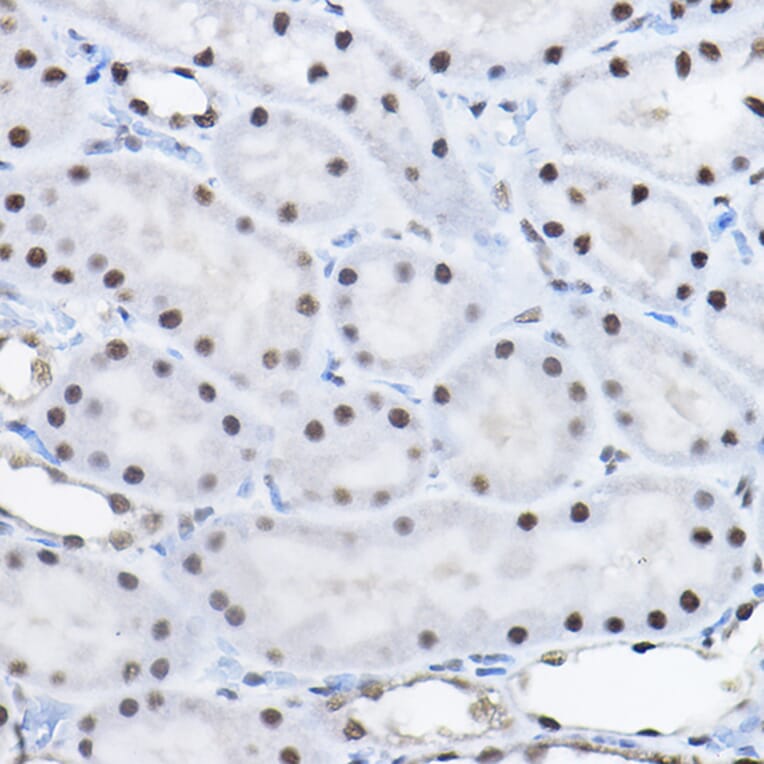
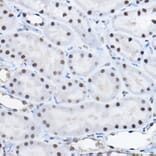
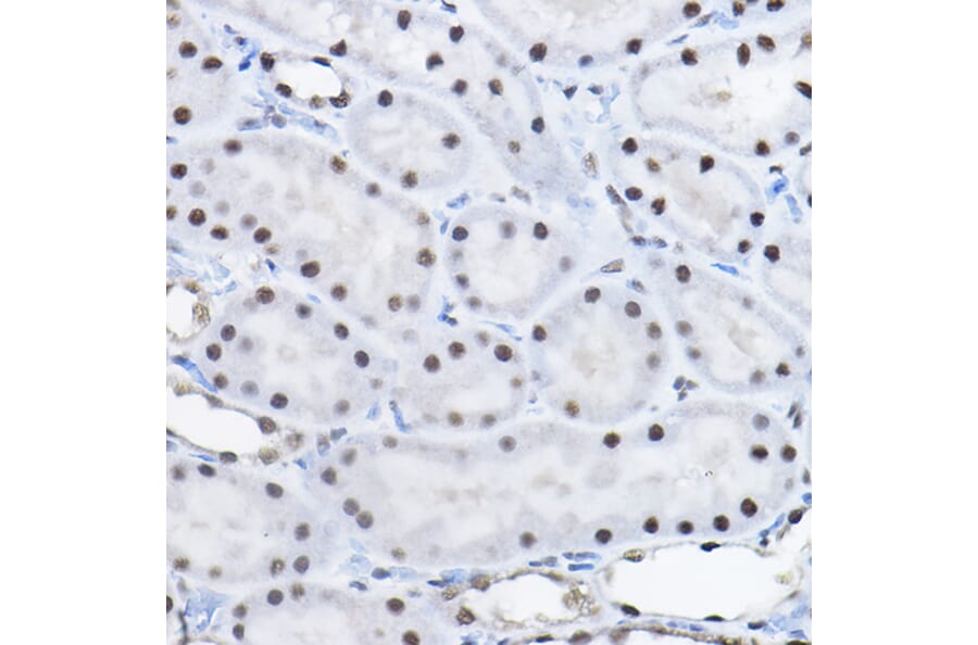
Immunohistochemistry analysis of paraffin-embedded rat kidney using Anti-BCAS2 Antibody (A14558) at a dilution of 1:100 (40x lens). Perform high pressure antigen retrieval with 10 mM citrate buffer pH 6.0 before commencing with IHC staining protocol.
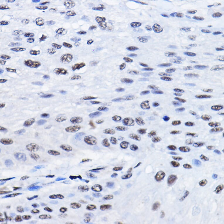
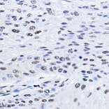
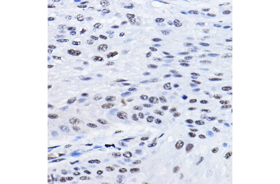
Immunohistochemistry analysis of paraffin-embedded human esophageal cancer using Anti-BCAS2 Antibody (A14558) at a dilution of 1:100 (40x lens). Perform high pressure antigen retrieval with 10 mM citrate buffer pH 6.0 before commencing with IHC staining protocol.
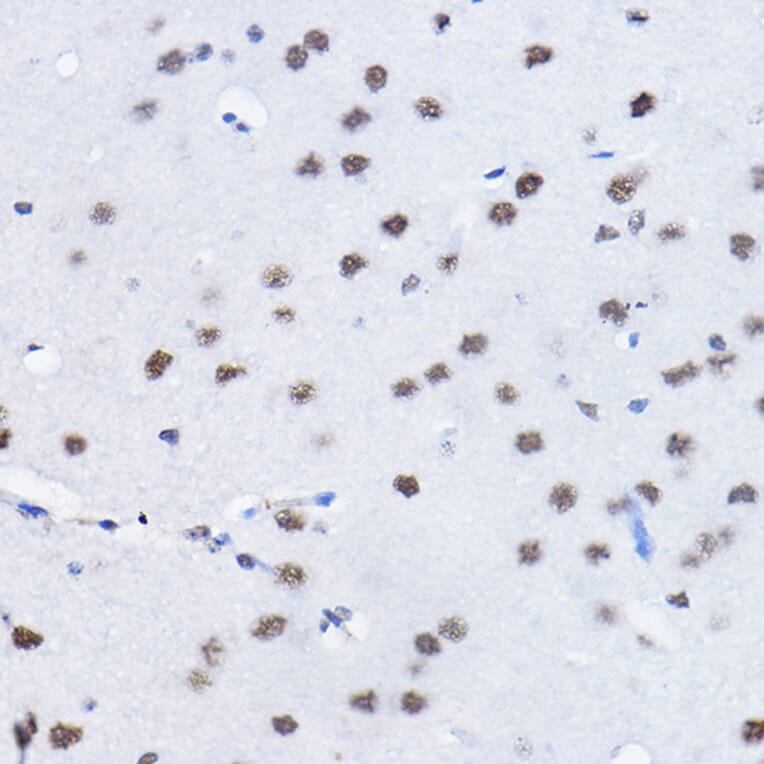
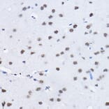
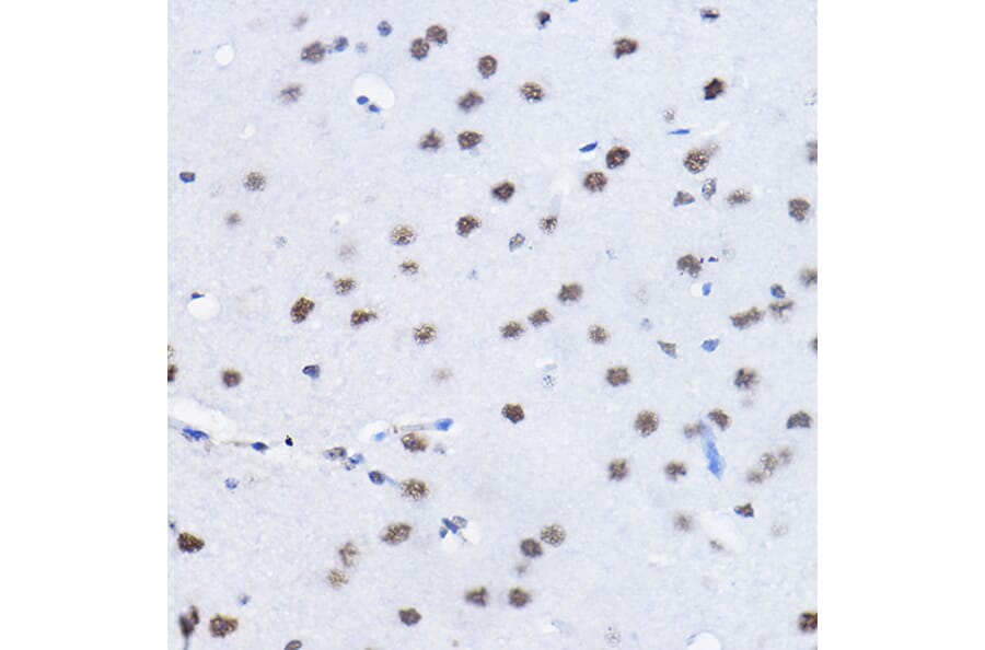
Immunohistochemistry analysis of paraffin-embedded mouse brain using Anti-BCAS2 Antibody (A14558) at a dilution of 1:100 (40x lens). Perform high pressure antigen retrieval with 10 mM citrate buffer pH 6.0 before commencing with IHC staining protocol.
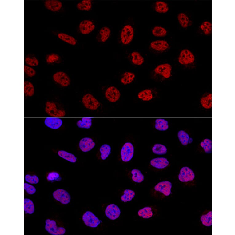
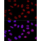
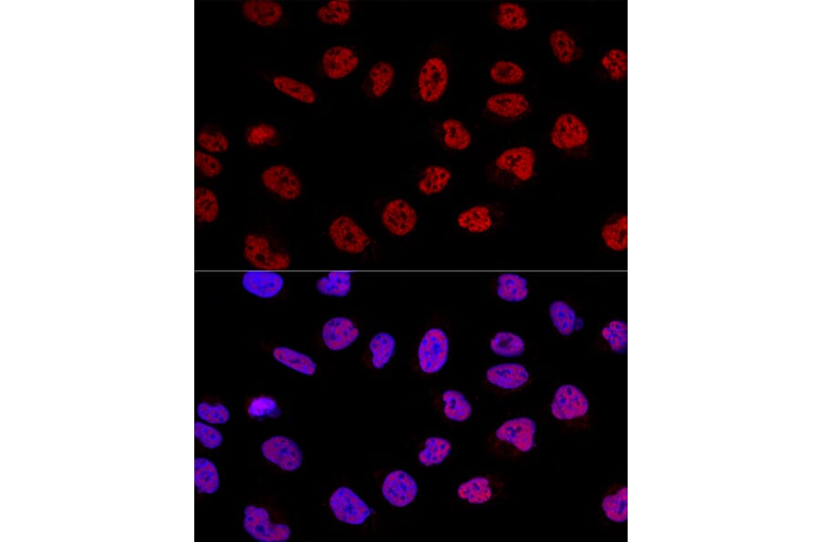
Confocal immunofluorescence analysis of U-2 OS cells using Anti-BCAS2 Antibody (A14558) at a dilution of 1:100. DAPI was used to stain the cell nuclei (blue).