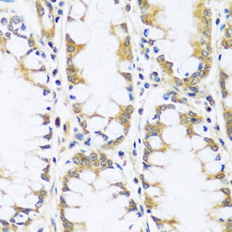
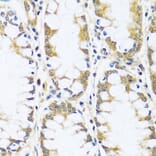
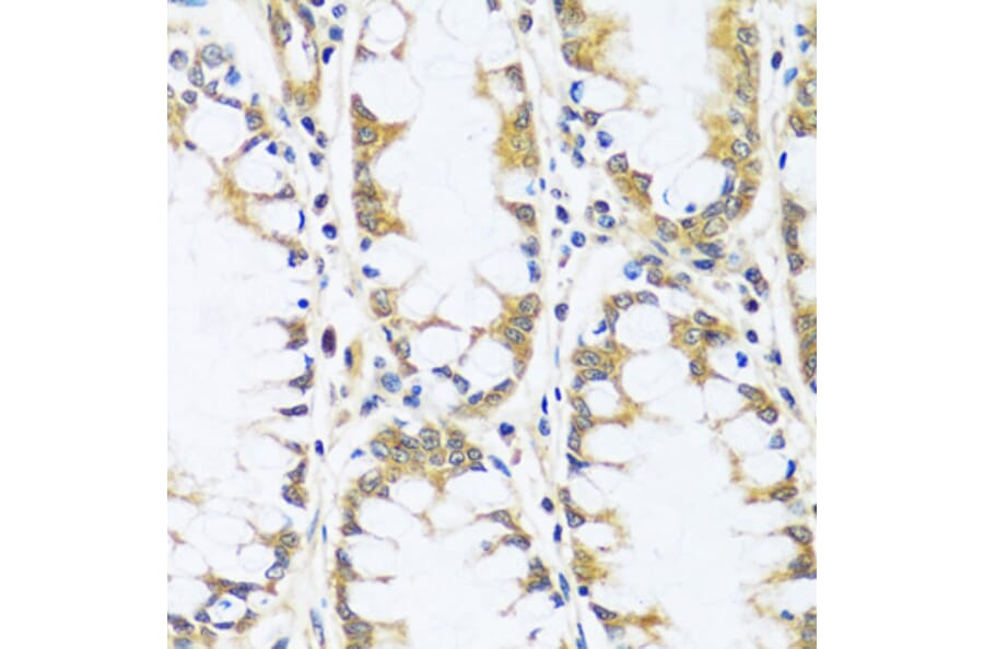
BLOC1S3 expression in human colon tissue analyzed by immunohistochemistry. Tissue was paraffin-embedded, and antigen retrieval was achieved by microwaving in 10 mM PBS buffer, pH 7.2. Staining was performed with Anti-BLOC1S3 Antibody (A92551) at a dilution of 1:100.
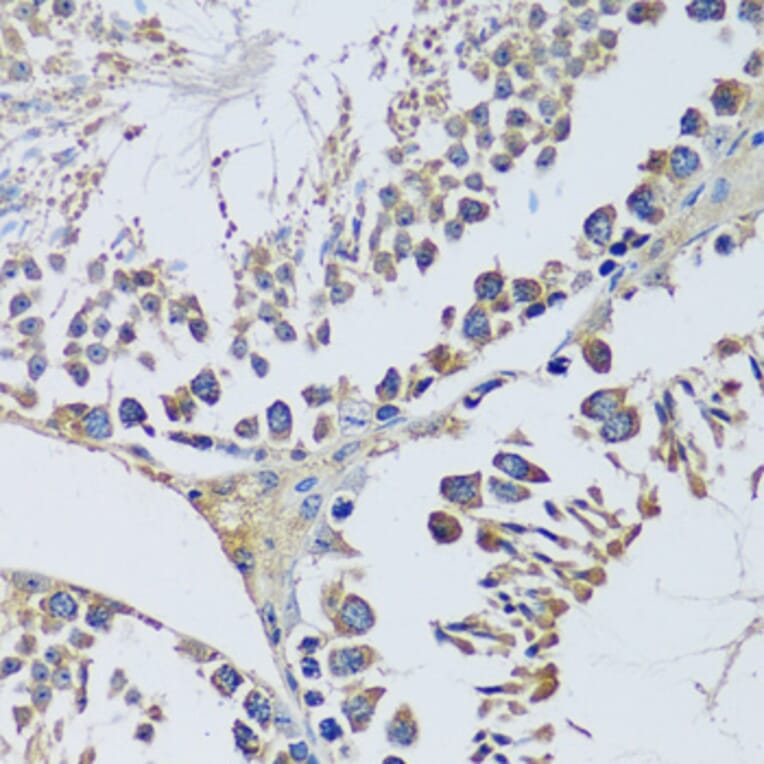
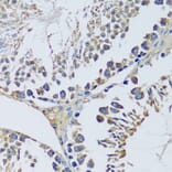
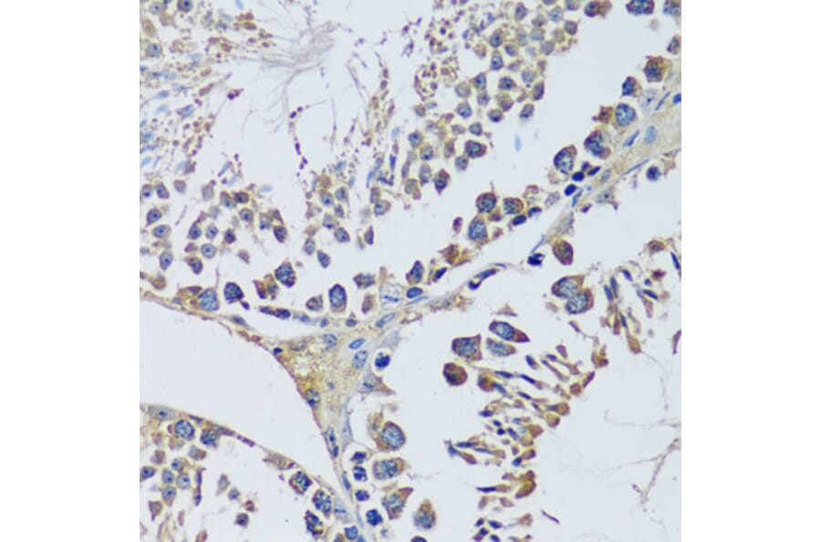
BLOC1S3 expression in mouse testis tissue analyzed by immunohistochemistry. Tissue was paraffin-embedded, and antigen retrieval was achieved by microwaving in 10 mM PBS buffer, pH 7.2. Staining was performed with Anti-BLOC1S3 Antibody (A92551) at a dilution of 1:100.
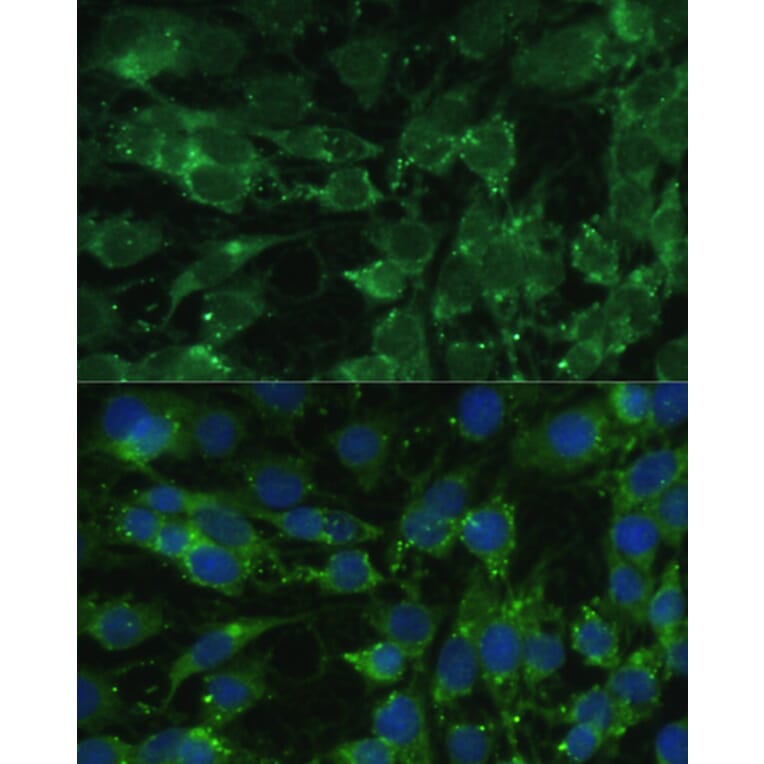
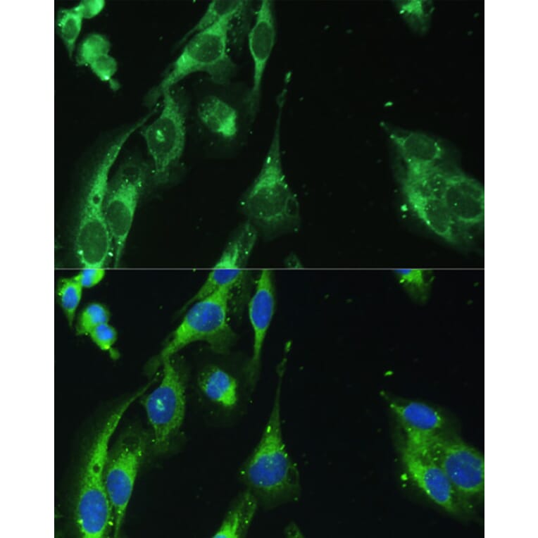
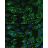
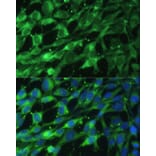
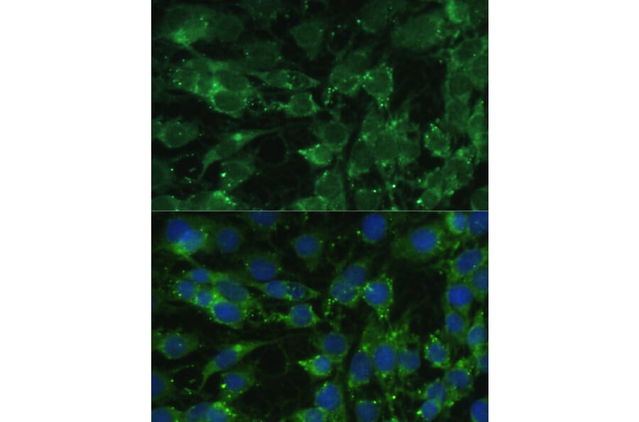
BLOC1S3 expression in C6 cells analyzed by immunofluorescence. Staining was performed with Anti-BLOC1S3 Antibody (A92551) at a dilution of 1:100 followed by Cy3 Goat Anti-Rabbit IgG (H+L) secondary antibody at a dilution of 1:500. Nuclei were stained with DAPI (blue).
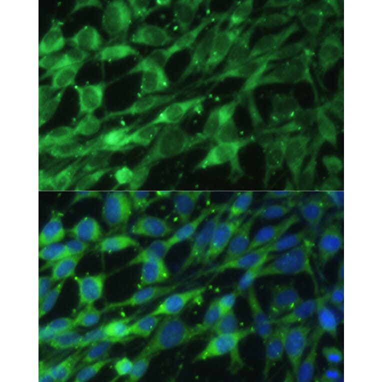
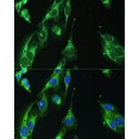
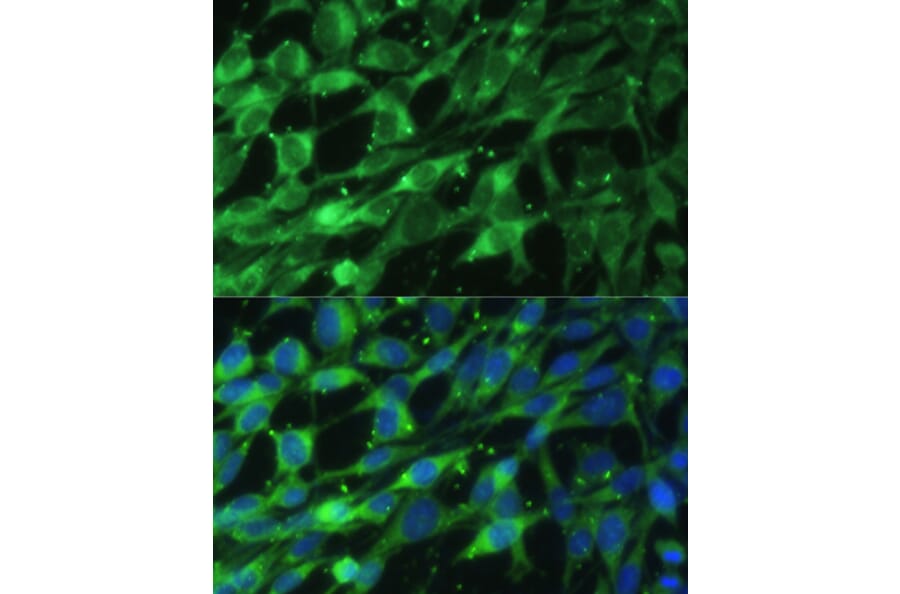
BLOC1S3 expression in NIH/3T3 cells analyzed by immunofluorescence. Staining was performed with Anti-BLOC1S3 Antibody (A92551) at a dilution of 1:100 followed by Cy3 Goat Anti-Rabbit IgG (H+L) secondary antibody at a dilution of 1:500. Nuclei were stained with DAPI (blue).
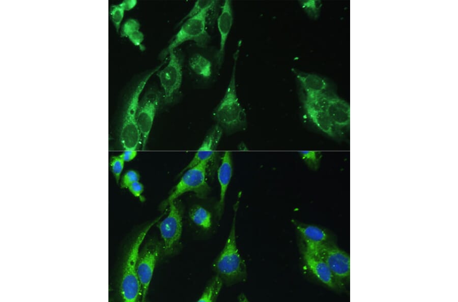
BLOC1S3 expression in U2OS cells analyzed by immunofluorescence. Staining was performed with Anti-BLOC1S3 Antibody (A92551) at a dilution of 1:100 followed by Cy3 Goat Anti-Rabbit IgG (H+L) secondary antibody at a dilution of 1:500. Nuclei were stained with DAPI (blue).