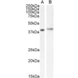
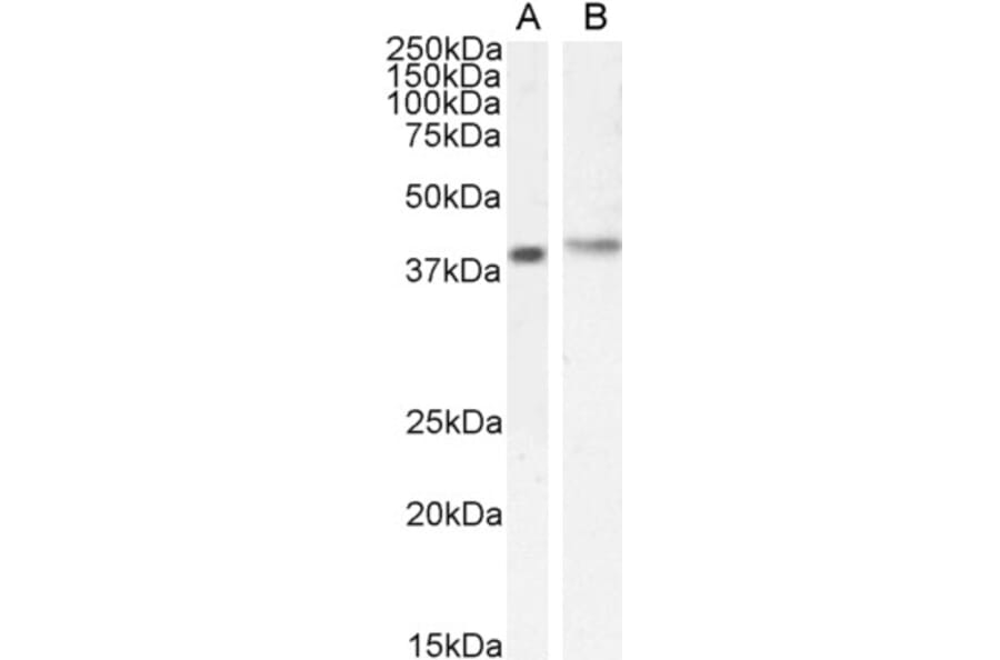
CASP expression in Jurkat (A) and MOLT4 (B) cell lysate analyzed by western blot. Cells were lysed in RIPA buffer and 35µg protein was run per lane. Primary incubation was performed with Anti-CASP Antibody (A83821) at 0.5µg/ml (A) or 1µg/ml (B) and detected by chemiluminescence.
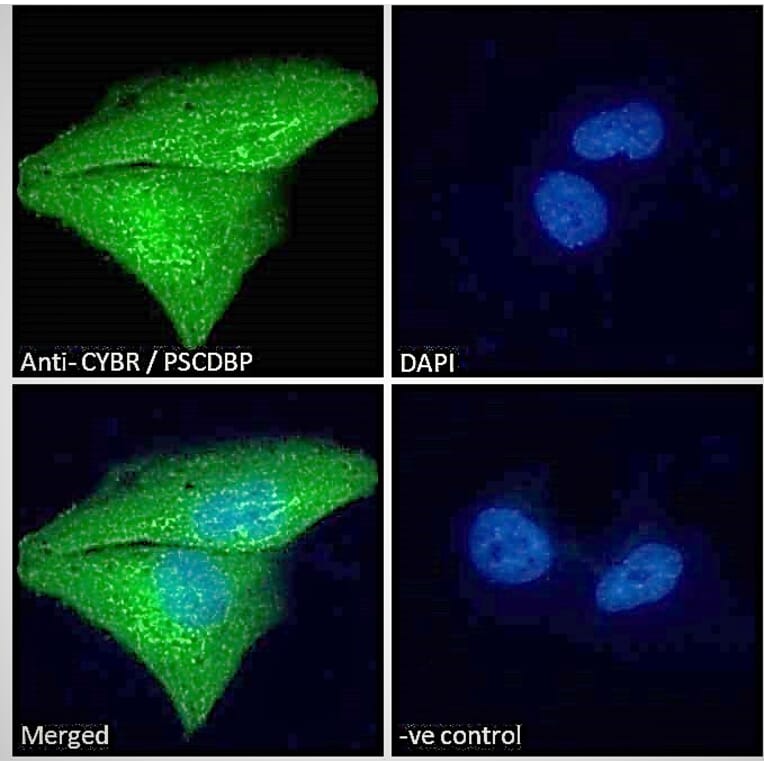
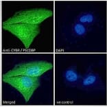
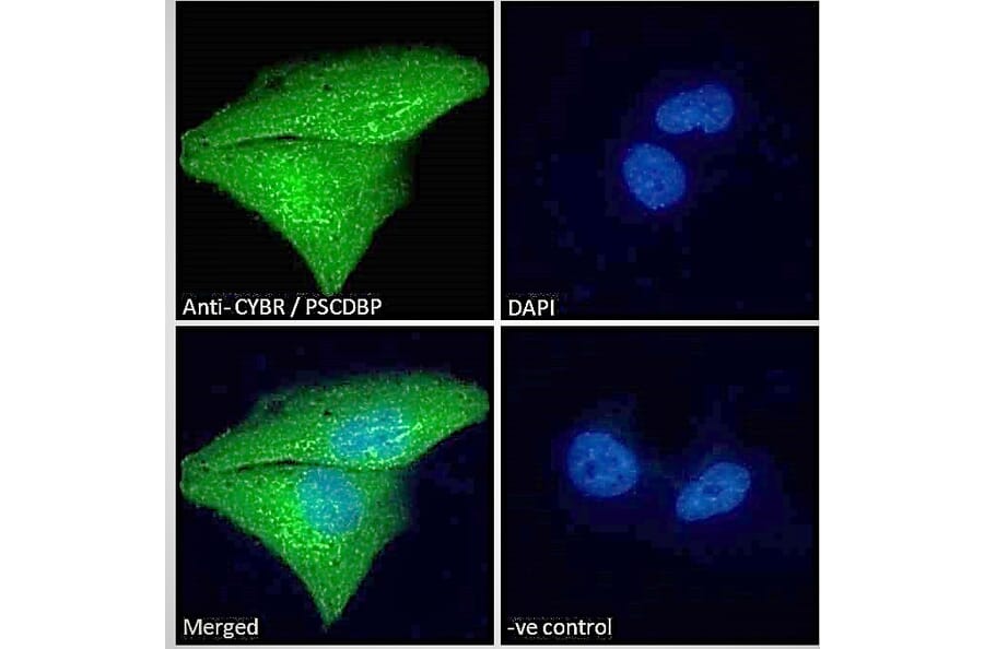
CASP expression in A431 cells analyzed by immunofluorescence. Cells were permeabilized with 0.15% Triton. Staining was performed with Anti-CASP Antibody (A83821) at 10µg/ml for 1 hour and Alexa Fluor 488 secondary antibody at 2µg/ml. Cytoplasmic and nuclear staining shown and nuclei were stained with DAPI (blue). Negative control: Goat IgG Isotype Control at 10µg/ml followed by Alexa Fluor 488 secondary antibody at 2µg/ml.
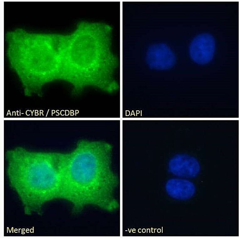
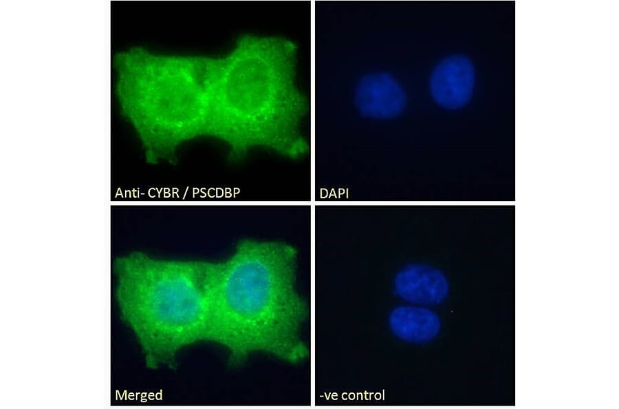
CASP expression in U251 cells analyzed by immunofluorescence. Cells were permeabilized with 0.15% Triton. Staining was performed with Anti-CASP Antibody (A83821) at 10µg/ml for 1 hour and Alexa Fluor 488 secondary antibody at 2µg/ml. Strong cytoplasmic and weak nuclear staining shown and nuclei were stained with DAPI (blue). Negative control: Goat IgG Isotype Control at 10µg/ml followed by Alexa Fluor 488 secondary antibody at 2µg/ml.
Publishing research using Anti-CASP Antibody (A83821)? Please let us know so that we can list the citation on this page.
Proteins predicted to interact with CASP
Predicted protein interactions based upon String database. Revelancy score correlates with probability of interaction.