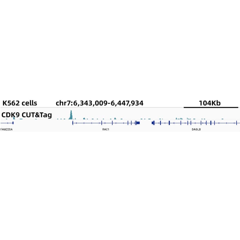
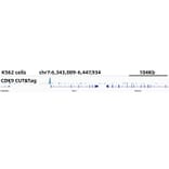
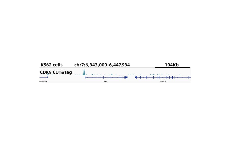
CUT&Tag was performed using the CUT&Tag Assay Kit (pAG-Tn5) for Illumina from 105 K562 cells with 5µg of Anti-Cdk9 Antibody (A81021), and Goat Anti-Rabbit IgG H&L Antibody. The CUT&Tag results indicate the enrichment pattern of CDK9 in representative gene loci (RAC1), as shown in figure.
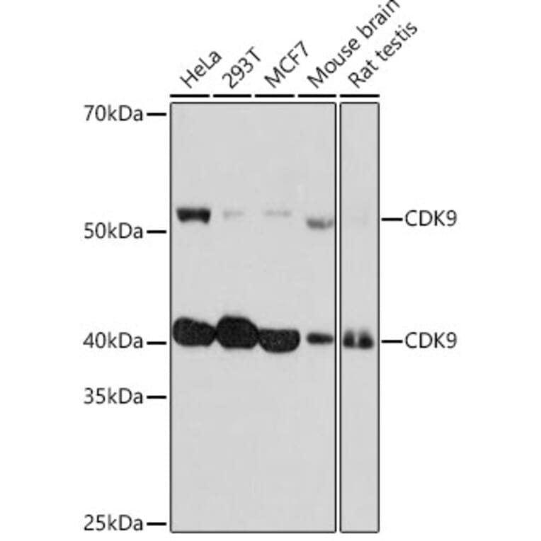
Western Blot - Anti-Cdk9 Antibody [ARC0527] (A81021)
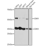
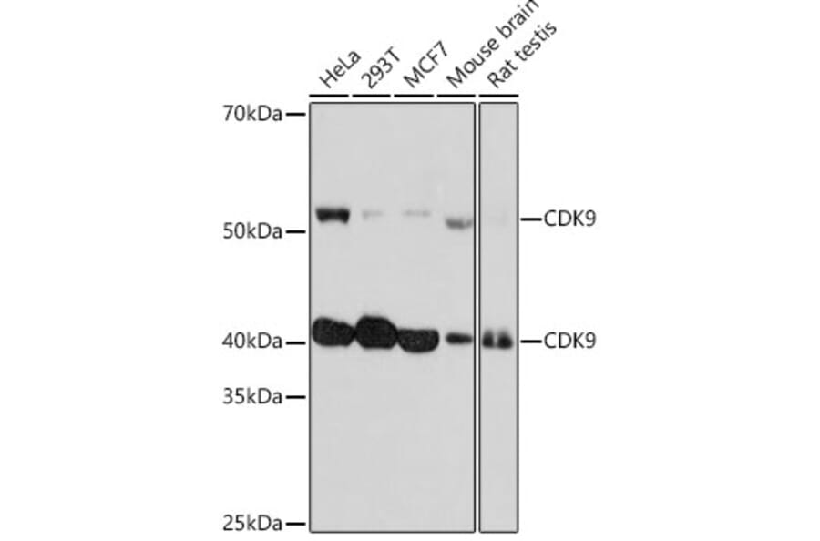
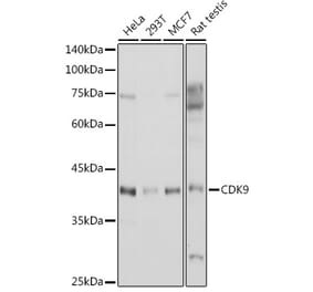
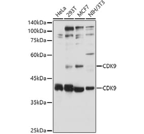
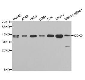
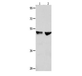
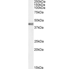
Western blot analysis of extracts of various cell lines, using Anti-Cdk9 Antibody (A81021) at 1:1,000 dilution. The secondary antibody was Goat Anti-Rabbit IgG H&L Antibody (HRP) at 1:10,000 dilution. Lysates/proteins were present at 25µg per lane. The blocking buffer used was 3% non-fat dry milk in TBST. Detection was with a ECL Basic Kit. Exposure time: 3min.
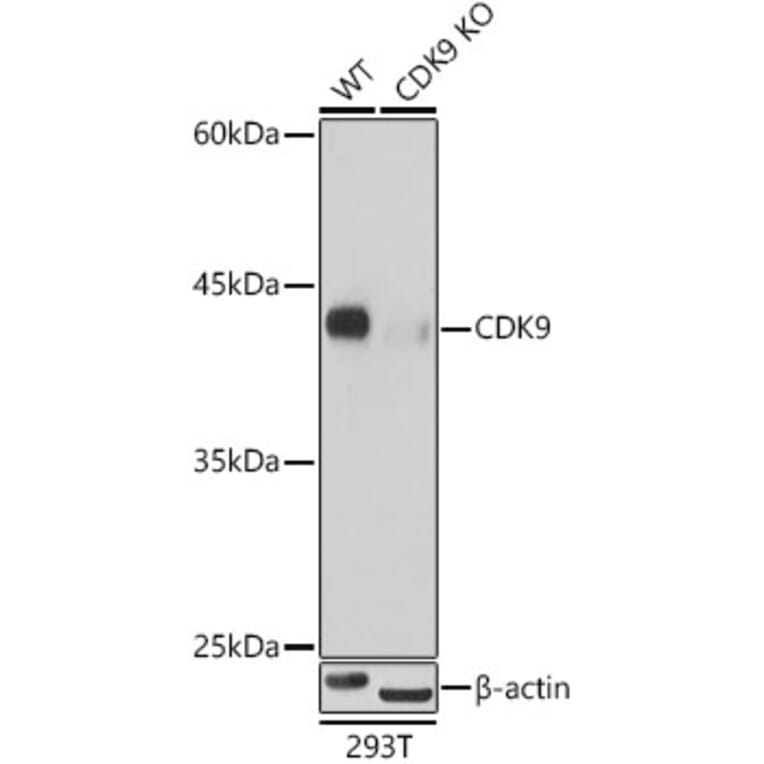
Western Blot - Anti-Cdk9 Antibody [ARC0527] (A81021)
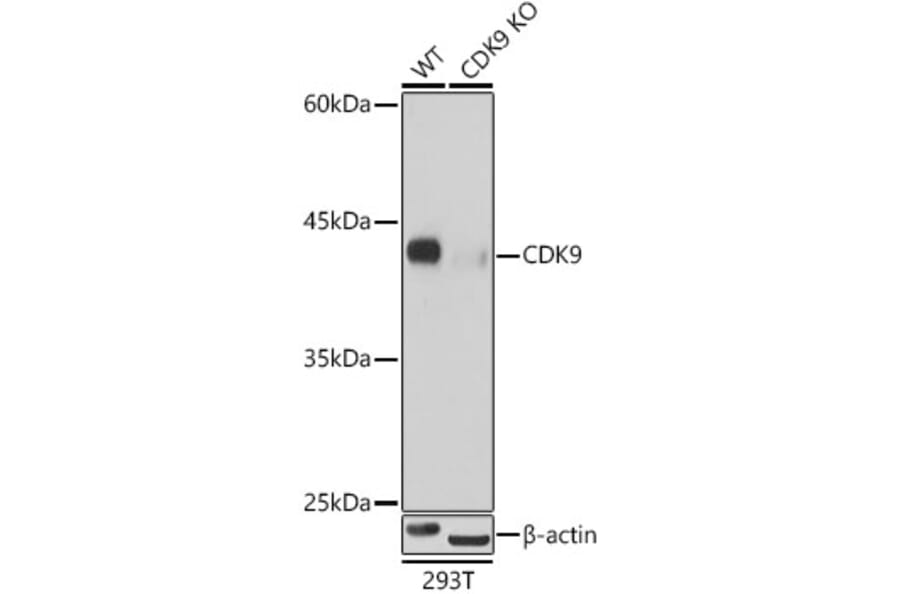
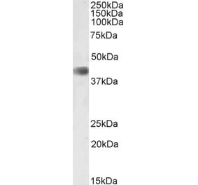
Western blot analysis of extracts from wild type(WT) and CDK9 knockout (KO) 293T cells, using Anti-Cdk9 Antibody (A81021) at 1:500 dilution. The secondary antibody was Goat Anti-Rabbit IgG H&L Antibody (HRP) at 1:10,000 dilution. Lysates/proteins were present at 25µg per lane. The blocking buffer used was 3% non-fat dry milk in TBST. Detection was with a ECL Basic Kit. Exposure time: 1s.
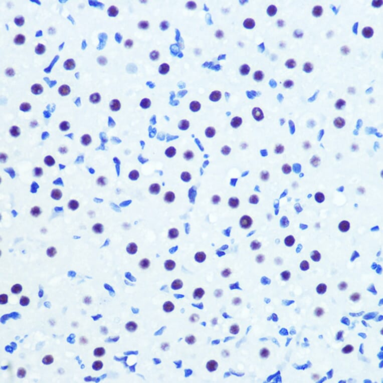
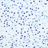
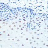
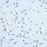
Immunohistochemistry analysis of paraffin-embedded rat ovary using Anti-Cdk9 Antibody (A81021) at a dilution of 1:100 (40x lens). Perform microwave antigen retrieval with 10 mM PBS buffer pH 7.2 before commencing with IHC staining protocol.
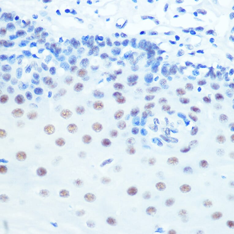
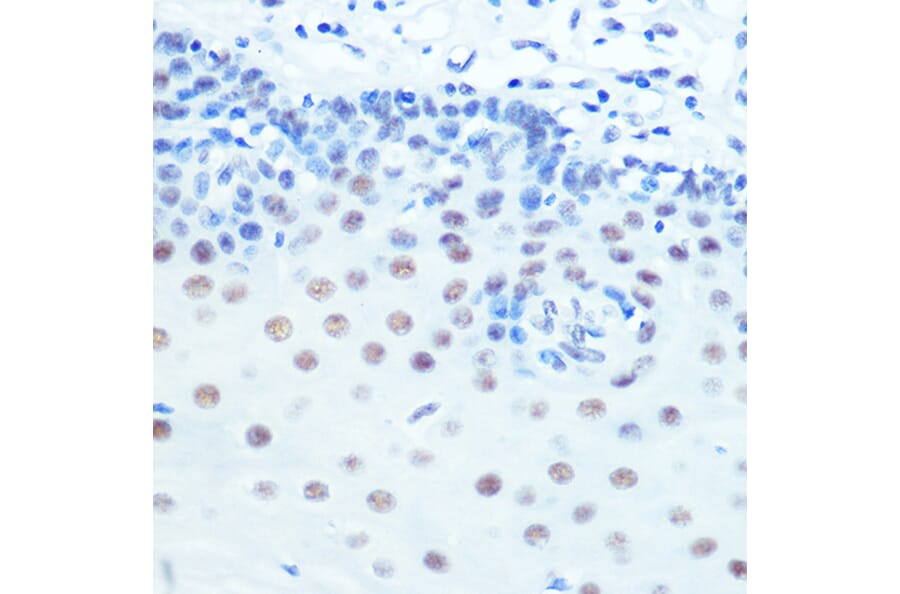
Immunohistochemistry analysis of paraffin-embedded human esophageal using Anti-Cdk9 Antibody (A81021) at a dilution of 1:100 (40x lens). Perform microwave antigen retrieval with 10 mM PBS buffer pH 7.2 before commencing with IHC staining protocol.
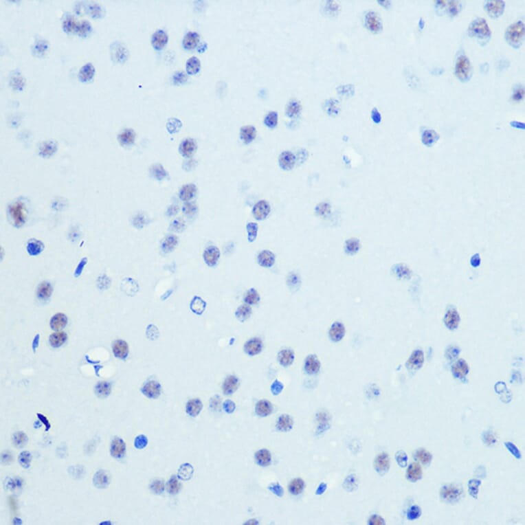
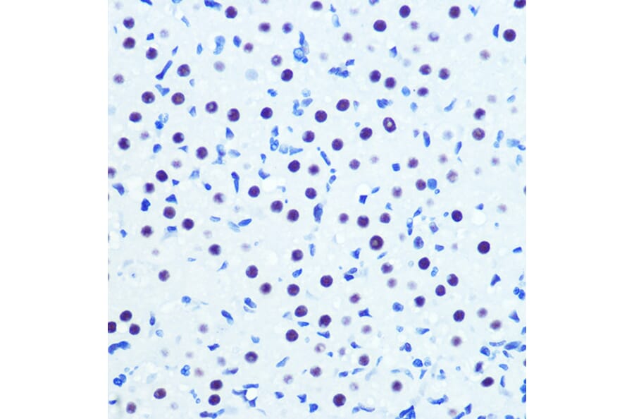
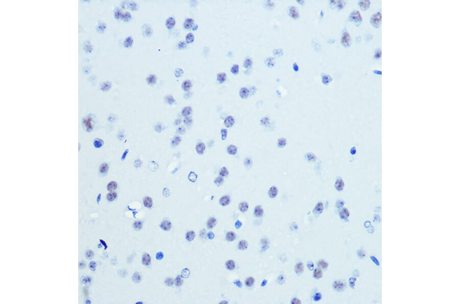
Immunohistochemistry analysis of paraffin-embedded mouse brain using Anti-Cdk9 Antibody (A81021) at a dilution of 1:100 (40x lens). Perform microwave antigen retrieval with 10 mM PBS buffer pH 7.2 before commencing with IHC staining protocol.
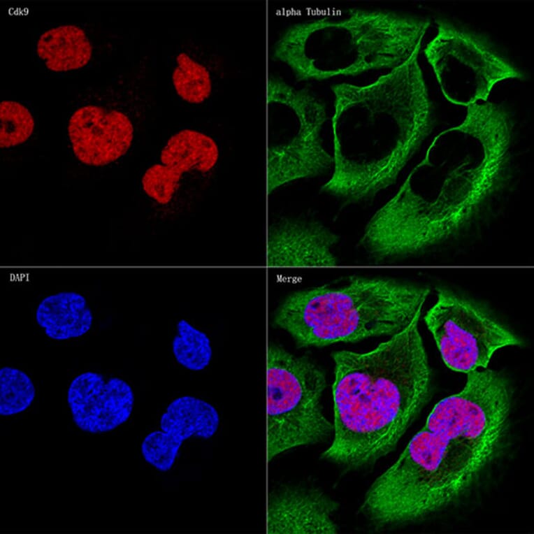
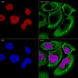
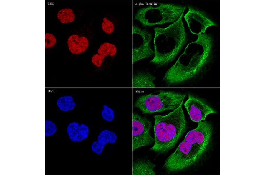
Confocal imaging of U-2 0S cells using Anti-Cdk9 Antibody (A81021), at a dilution of 1:50, (red). The cells were counterstained with Anti-alpha Tubulin Antibody, at a dilution of 1:400, (green). DAPI was used for nuclear staining (Blue). Objective: 60x.
Western Blot - Anti-Cdk9 Antibody [ARC0527] (A81021)
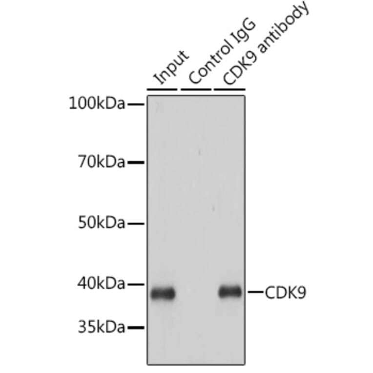
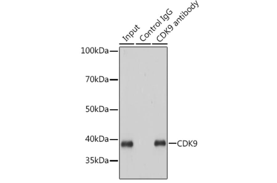
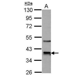
Immunoprecipitation analysis of 200µg extracts of HeLa cells using 3µg of Anti-Cdk9 Antibody (A81021). This Western blot was performed on the immunoprecipitate using Anti-Cdk9 Antibody (A81021) at a dilution of 1:1000.
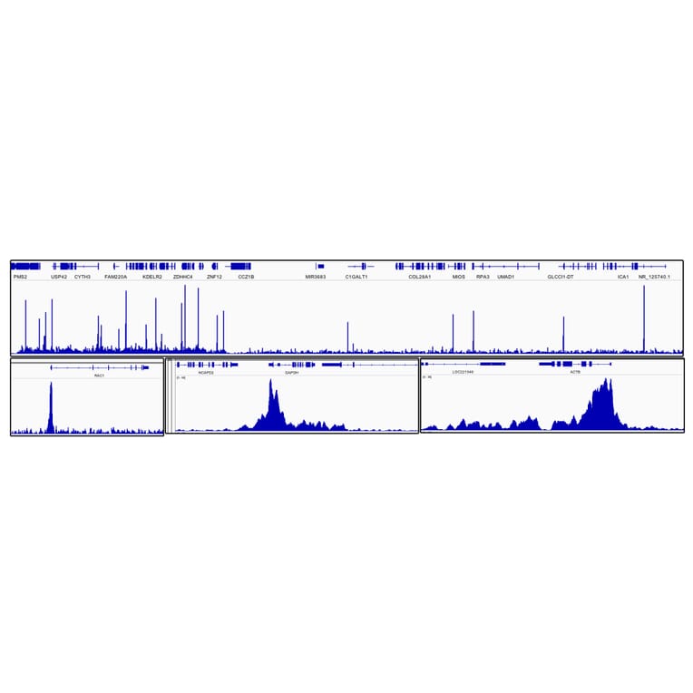
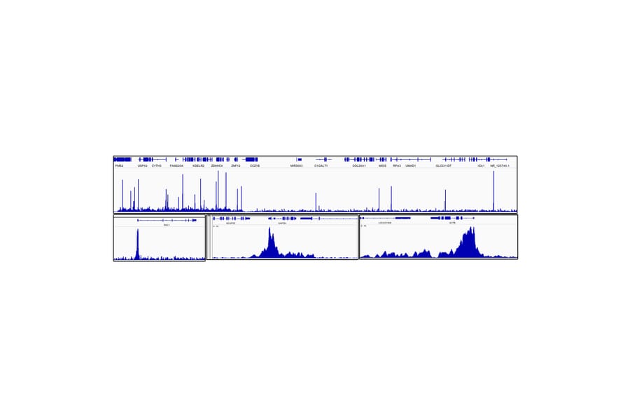
Chromatin immunoprecipitations analysis of cross-linked chromatin from DLD-1 cells, using Anti-Cdk9 Antibody (A81021). The ChIP sequencing results indicate the enrichment pattern of CDK9 in selected genomic region and representative gene loci (e. g. RAC1, GAPDH and ACTB), as shown in figure.