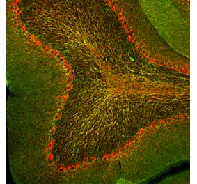
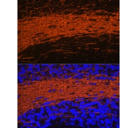
Immunofluorescent analysis of rat cerebellum section stained with Anti-CNPase Antibody, at a dilution of 1:500, in green, and Anti-NF-M Antibody (A85324 | 1:1,000, in red. The blue is DAPI staining of nuclear DNA. Following transcardial perfusion of rat with 4% paraformaldehyde, brain was post fixed for 24 hours, cut to 45 µM, and free-floating sections were stained with the above antibodies. The Anti-CNPase Antibody stains the oligodendrocytes, cells that create the myelin sheath around axons. Anti-NF-M Antibody labels axons of neuronal cells.
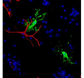
Mixed neuron-glial cell cultures stained with Anti-CNPase Antibody (red) and Anti-GFAP Antibody (A85419 | green). The Anti-CNPase Antibody stains strongly in oligodendrocytes, whereas Anti-GFAP Antibody labels only the intermediate filaments in astrocytes. Blue is DNA staining.
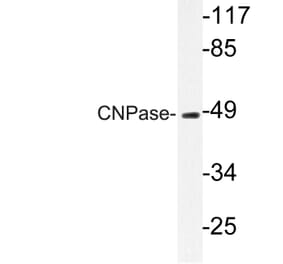
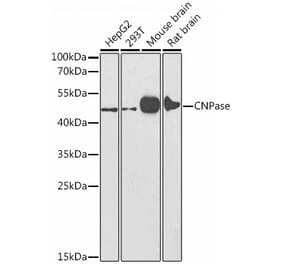
Western Blot - Anti-CNPase Antibody [1H10] (A85413)
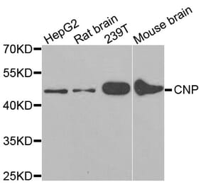
Western blot analysis of different tissue lysates using Anti-CNPase Antibody, at a dilution of 1:2,000, in green,: [Lane 1] protein standard (red), [Lane 2] rat brain, [Lane 3] rat spinal cord, [Lane 4] mouse brain, [Lane 5] mouse spinal cord. Double bands at 46, 48 kDa mark correspond to isotypes of the CNP protein.
Immunohistochemistry analysis of a 4%PFA fixed paraffin embedded rat cerebellum section with Anti-CNPase Antibody [1H10] (A85413) at a dilution of 1:1,000 detected in DAB (brown) following the Vector Labs mouse on mouse Immpress method and reagents with citra buffer retrieval. Counterstained with Hematoxylin (blue). In the cerebellum, the CNP antibody labels white matter cells, processes in the white matter and processes in the granular layer. Note: this antibody performs well in testing with both 4% PFA and standard NBF fixed tissues.
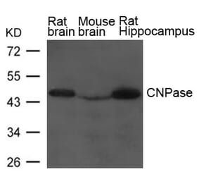
Western Blot - Anti-CNPase Antibody [1H10] (A85413)
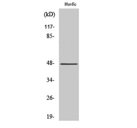
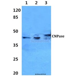
Blots of rat brain tissue homogenates probed with Anti-CNPase Antibody at 1:5,000 (Lane 1) and 1:20,000 (Lane 2). The antibody binds strongly and cleanly to a band at ~48kDa.
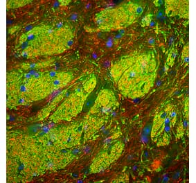
Immunofluorescent analysis of rat cortex section stained with Anti-Adenylate Cyclase 3 Antibody (A104341), at a dilution of 1:10,000, in red, and co-stained with Anti-CNPase Antibody [1H10] (A85413), at a dilution of 1:1,000, in green. The blue is Hoechst staining of nuclear DNA. Anti-Adenylate Cyclase 3 Antibody (A104341) reveals neuronal cilia while Anti-CNPase Antibody [1H10] (A85413) stains oligodendrocytes and the myelin sheath around axons.


![Immunofluorescence - Anti-CNPase Antibody [1H10] (A85413) - Antibodies.com](https://cdn.antibodies.com/image/catalog/85/A85413_1.jpg?profile=product_top)
![Immunofluorescence - Anti-CNPase Antibody [1H10] (A85413) - Antibodies.com](https://cdn.antibodies.com/image/catalog/85/A85413_2.jpg?profile=product_top)
![Western Blot - Anti-CNPase Antibody [1H10] (A85413) - Antibodies.com](https://cdn.antibodies.com/image/catalog/85/A85413_3.jpg?profile=product_top)
![Immunohistochemistry - Anti-CNPase Antibody [1H10] (A85413) - Antibodies.com](https://cdn.antibodies.com/image/catalog/85/A85413_4.jpg?profile=product_top)
![Western Blot - Anti-CNPase Antibody [1H10] (A85413) - Antibodies.com](https://cdn.antibodies.com/image/catalog/85/A85413_5.jpg?profile=product_top)
![Immunofluorescence - Anti-CNPase Antibody [1H10] (A85413) - Antibodies.com](https://cdn.antibodies.com/image/catalog/85/A85413_6.jpg?profile=product_top)
![Immunofluorescence - Anti-CNPase Antibody [1H10] (A85413) - Antibodies.com](https://cdn.antibodies.com/image/catalog/85/A85413_1.jpg?profile=product_top_thumb)
![Immunofluorescence - Anti-CNPase Antibody [1H10] (A85413) - Antibodies.com](https://cdn.antibodies.com/image/catalog/85/A85413_2.jpg?profile=product_top_thumb)
![Western Blot - Anti-CNPase Antibody [1H10] (A85413) - Antibodies.com](https://cdn.antibodies.com/image/catalog/85/A85413_3.jpg?profile=product_top_thumb)
![Immunohistochemistry - Anti-CNPase Antibody [1H10] (A85413) - Antibodies.com](https://cdn.antibodies.com/image/catalog/85/A85413_4.jpg?profile=product_top_thumb)
![Western Blot - Anti-CNPase Antibody [1H10] (A85413) - Antibodies.com](https://cdn.antibodies.com/image/catalog/85/A85413_5.jpg?profile=product_top_thumb)
![Immunofluorescence - Anti-CNPase Antibody [1H10] (A85413) - Antibodies.com](https://cdn.antibodies.com/image/catalog/85/A85413_6.jpg?profile=product_top_thumb)
![Immunofluorescence - Anti-CNPase Antibody [1H10] (A85413) - Antibodies.com](https://cdn.antibodies.com/image/catalog/85/A85413_1.jpg?profile=product_image)
![Immunofluorescence - Anti-CNPase Antibody [1H10] (A85413) - Antibodies.com](https://cdn.antibodies.com/image/catalog/85/A85413_2.jpg?profile=product_image)
![Western Blot - Anti-CNPase Antibody [1H10] (A85413) - Antibodies.com](https://cdn.antibodies.com/image/catalog/85/A85413_3.jpg?profile=product_image)
![Immunohistochemistry - Anti-CNPase Antibody [1H10] (A85413) - Antibodies.com](https://cdn.antibodies.com/image/catalog/85/A85413_4.jpg?profile=product_image)
![Western Blot - Anti-CNPase Antibody [1H10] (A85413) - Antibodies.com](https://cdn.antibodies.com/image/catalog/85/A85413_5.jpg?profile=product_image)
![Immunofluorescence - Anti-CNPase Antibody [1H10] (A85413) - Antibodies.com](https://cdn.antibodies.com/image/catalog/85/A85413_6.jpg?profile=product_image)