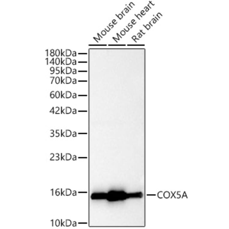
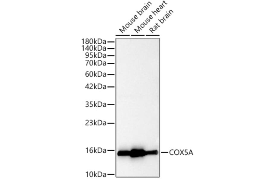
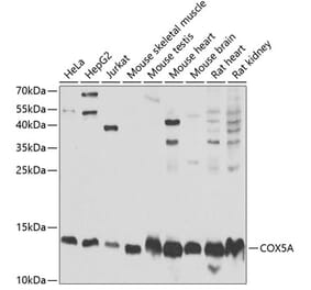
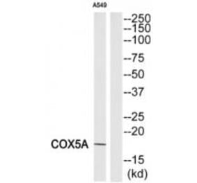
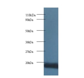
COX5A expression in various lysates analyzed by western blot. Primary antibody incubation was performed for 1 hour on 25ug protein per lane with Anti-COX5A Antibody (A329278) at a dilution of 1:2000 and detected with chemiluminescence.
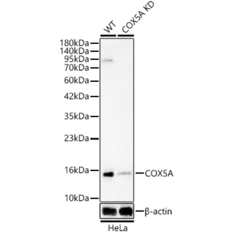
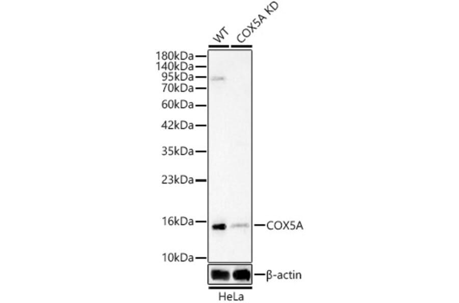
COX5A expression in lysates from wild type (WT) and COX5A knockdown (KD) HeLa(KD) cells analyzed by western blot. Primary antibody incubation was performed for 1 hour on 25ug protein per lane with Anti-COX5A Antibody (A329278) at a dilution of 1:2000 and detected with chemiluminescence.
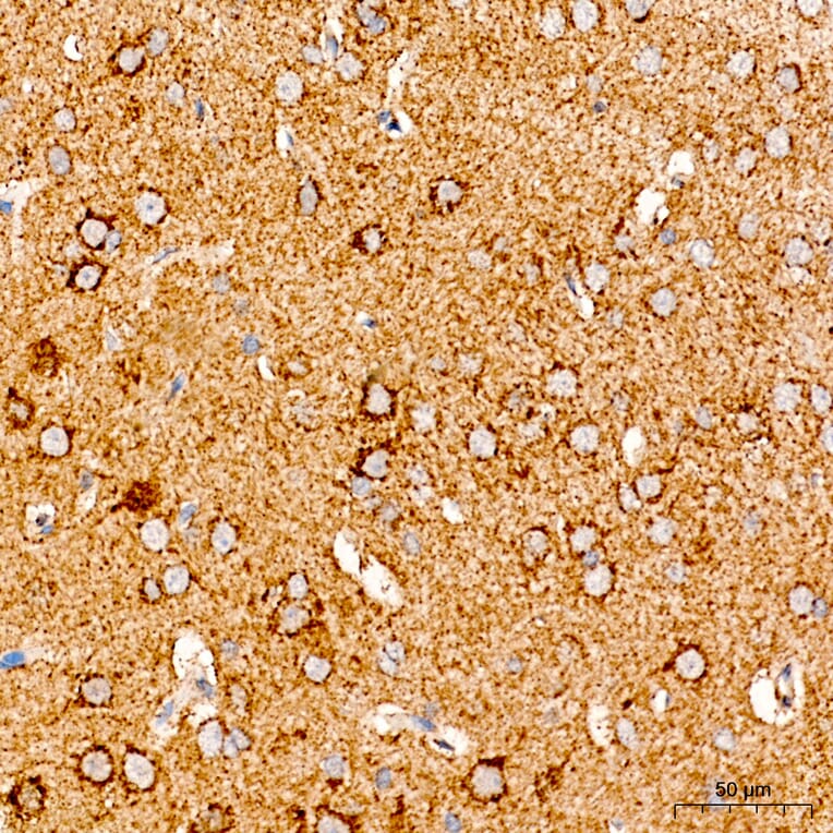
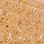
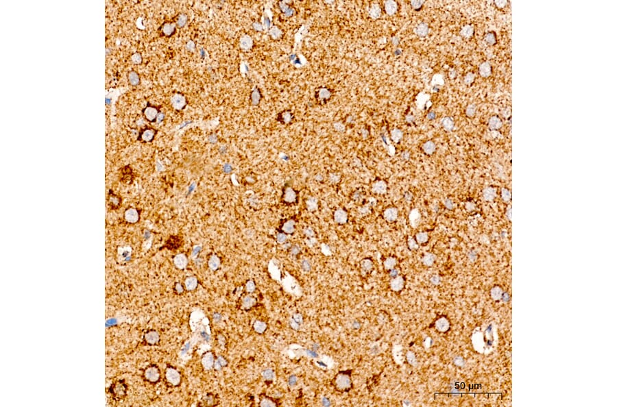
COX5A expression in rat brain tissue analyzed by immunohistochemistry. Tissue was paraffin-embedded, and antigen retrieval was achieved with 10 mM citrate buffer, pH 6.0, under high pressure. Staining was performed with Anti-COX5A Antibody (A329278) at a dilution of 1:100.
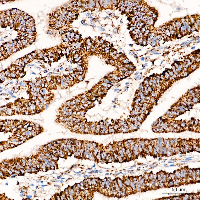
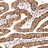
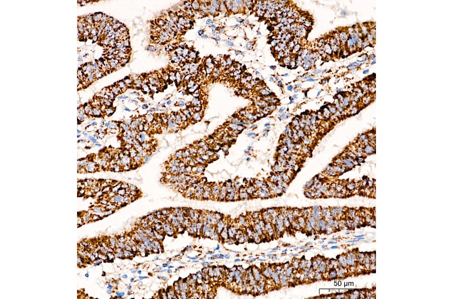
COX5A expression in human colon carcinoma tissue analyzed by immunohistochemistry. Tissue was paraffin-embedded, and antigen retrieval was achieved with 10 mM citrate buffer, pH 6.0, under high pressure. Staining was performed with Anti-COX5A Antibody (A329278) at a dilution of 1:100.
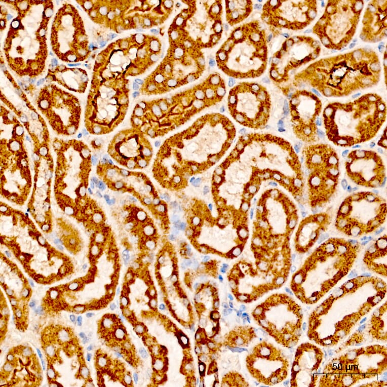
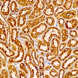
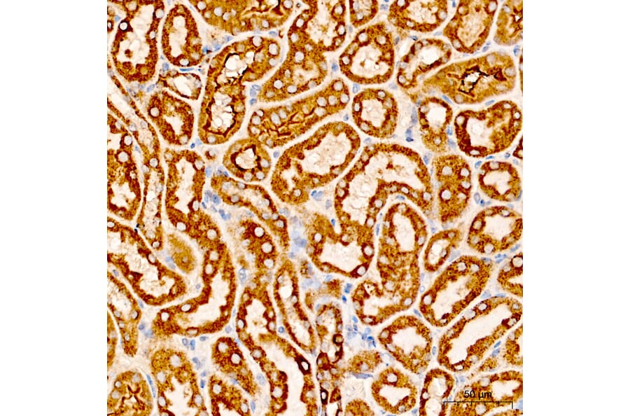
COX5A expression in mouse kidney tissue analyzed by immunohistochemistry. Tissue was paraffin-embedded, and antigen retrieval was achieved with 10 mM citrate buffer, pH 6.0, under high pressure. Staining was performed with Anti-COX5A Antibody (A329278) at a dilution of 1:100.
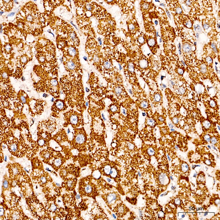
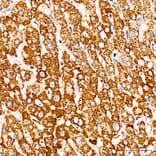
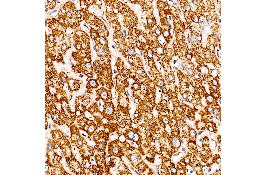
COX5A expression in human liver cancer tissue analyzed by immunohistochemistry. Tissue was paraffin-embedded, and antigen retrieval was achieved with 10 mM citrate buffer, pH 6.0, under high pressure. Staining was performed with Anti-COX5A Antibody (A329278) at a dilution of 1:100.
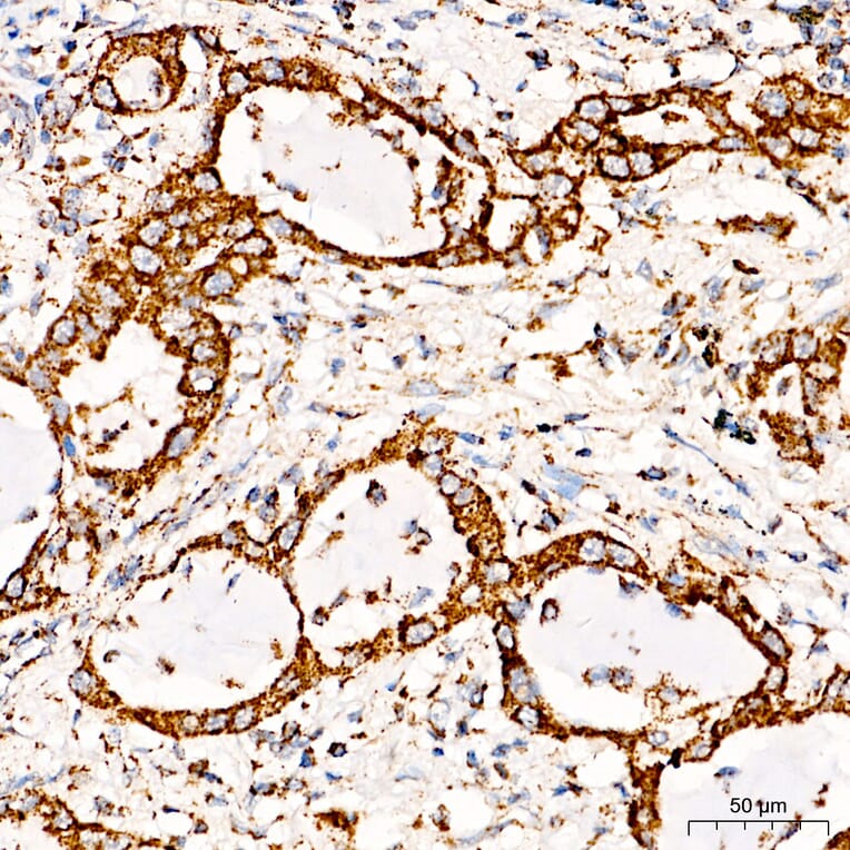
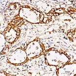
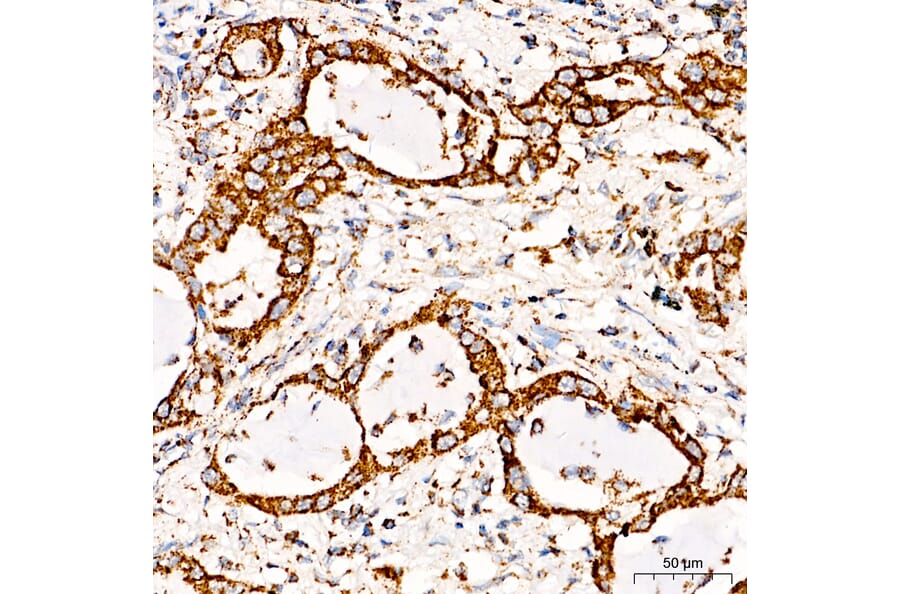
COX5A expression in human lung cancer tissue analyzed by immunohistochemistry. Tissue was paraffin-embedded, and antigen retrieval was achieved with 10 mM citrate buffer, pH 6.0, under high pressure. Staining was performed with Anti-COX5A Antibody (A329278) at a dilution of 1:100.