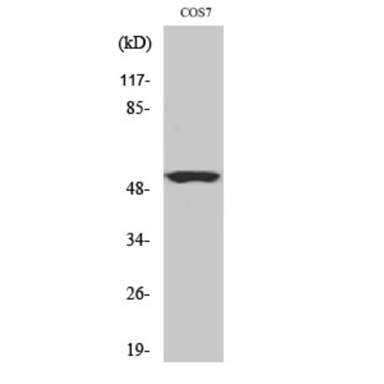
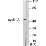
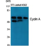
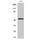
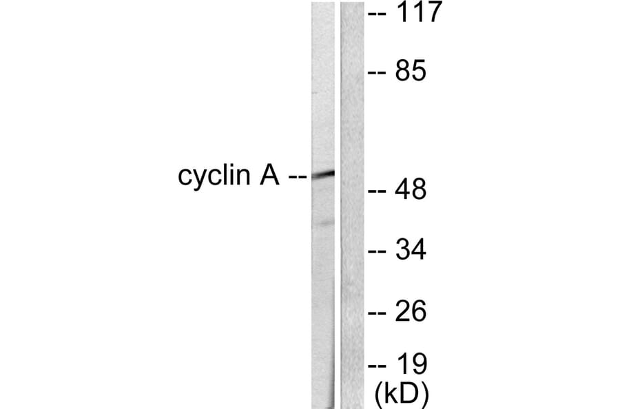
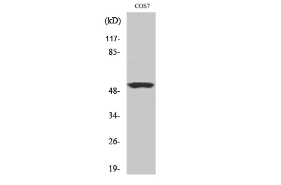
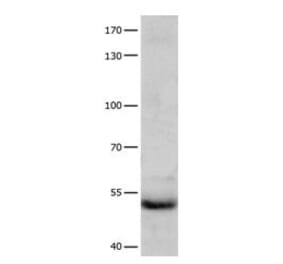
Western blot analysis of lysates from COS7 cells using Anti-Cyclin A Antibody. The right hand lane represents a negative control, where the antibody is blocked by the immunising peptide.
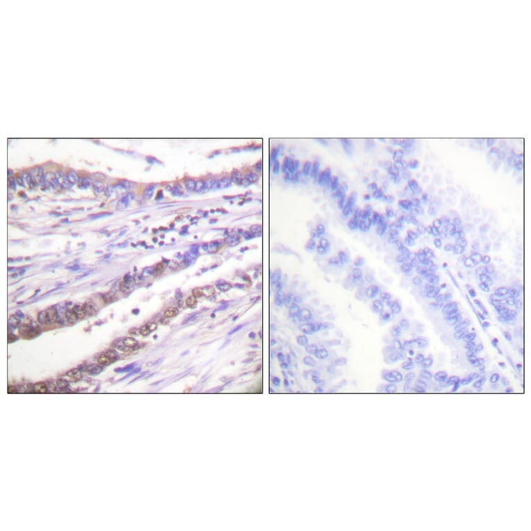
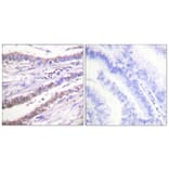
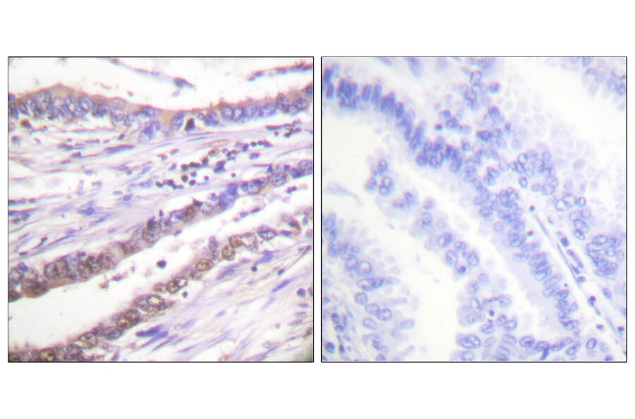
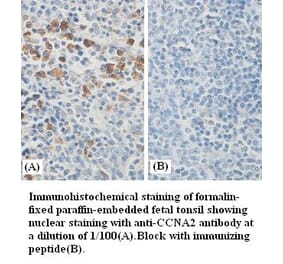
Immunohistochemistry - Anti-Cyclin A Antibody (A94878)
Immunohistochemical analysis of paraffin-embedded human lung carcinoma tissue using Anti-Cyclin A Antibody. The right hand panel represents a negative control, where the antibody was pre-incubated with the immunising peptide.
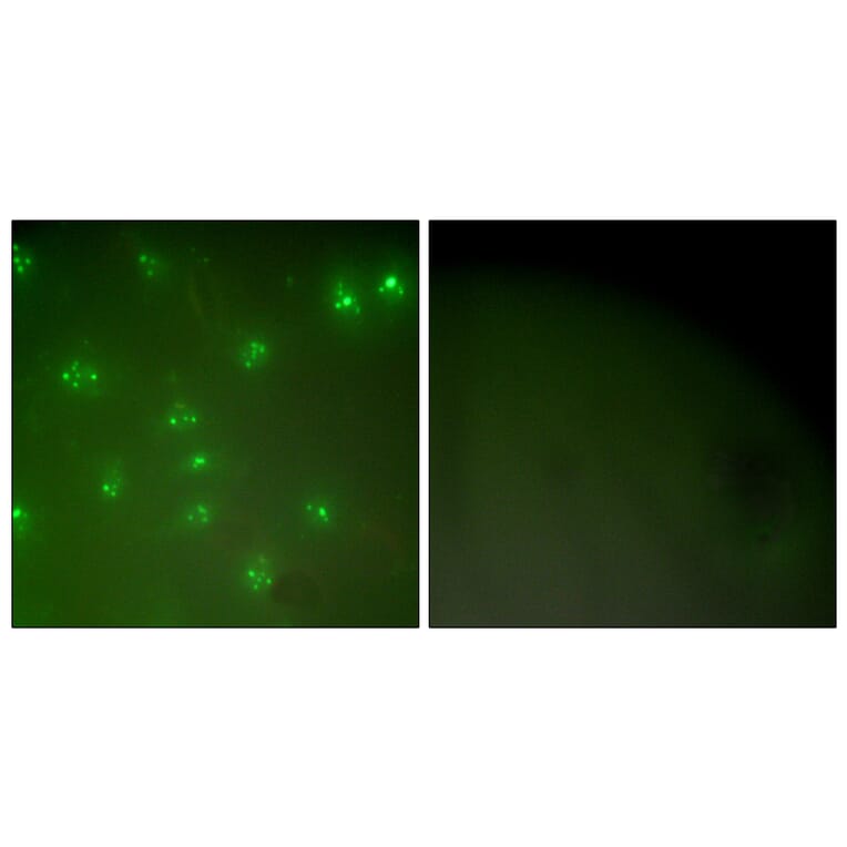
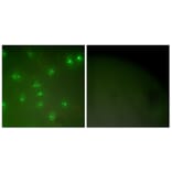
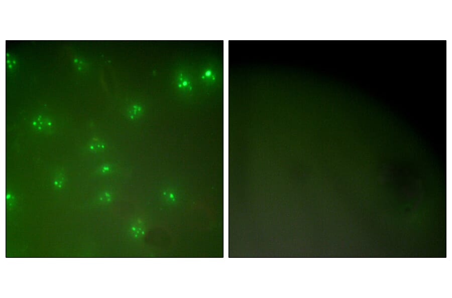
Immunofluorescence - Anti-Cyclin A Antibody (A94878)
Immunofluorescence analysis of COS7 cells using Anti-Cyclin A Antibody. The right hand panel represents a negative control, where the antibody was pre-incubated with the immunising peptide.
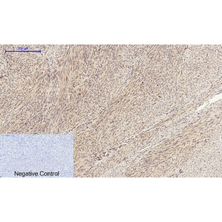
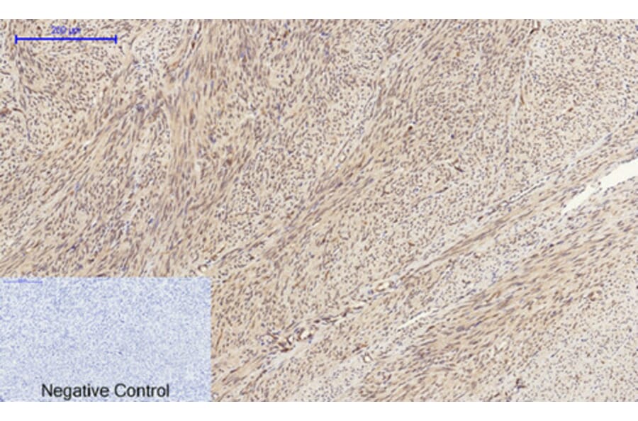
Immunohistochemistry - Anti-Cyclin A Antibody (A94878)
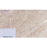
Immunohistochemical analysis of paraffin-embedded human uterus tissue using Anti-Cyclin A Antibody at 1:200 (4°C overnight). Negative control was secondary antibody only.
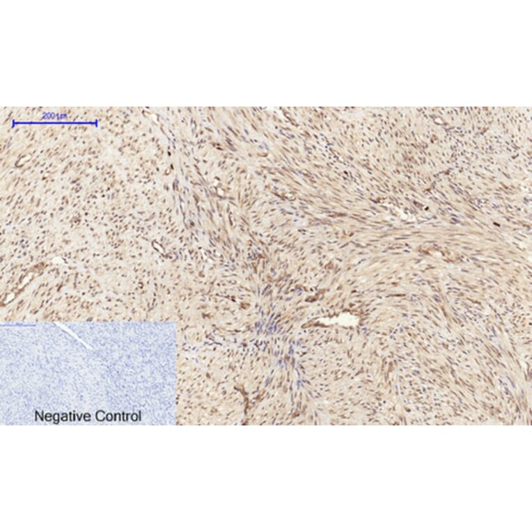
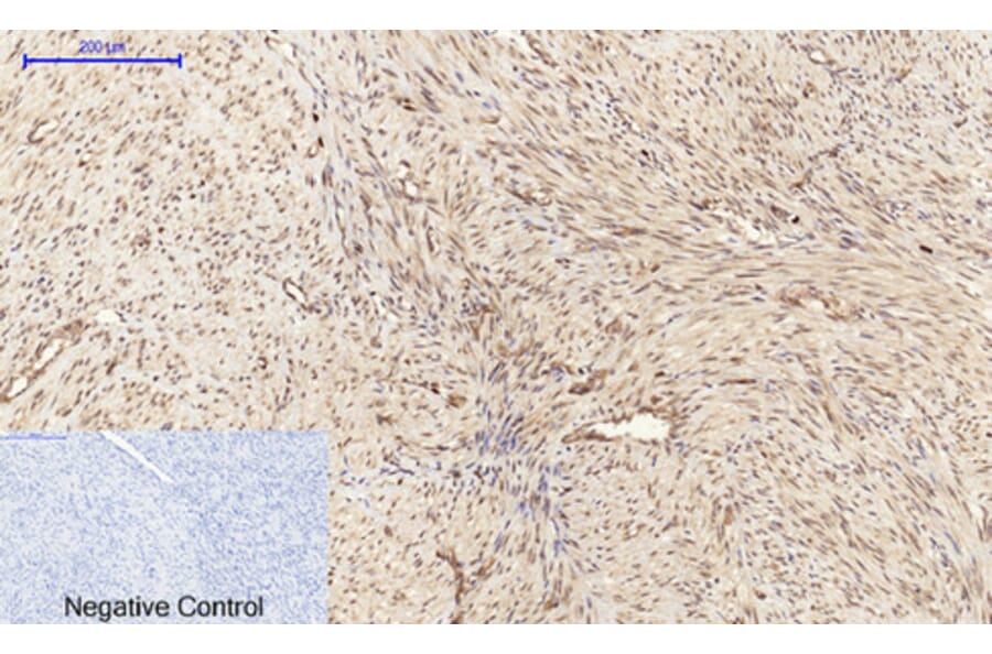
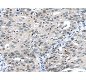
Immunohistochemistry - Anti-Cyclin A Antibody (A94878)
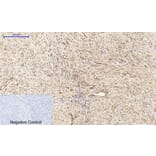
Immunohistochemical analysis of paraffin-embedded human uterus cancer tissue using Anti-Cyclin A Antibody at 1:200 (4°C overnight). Negative control was secondary antibody only.
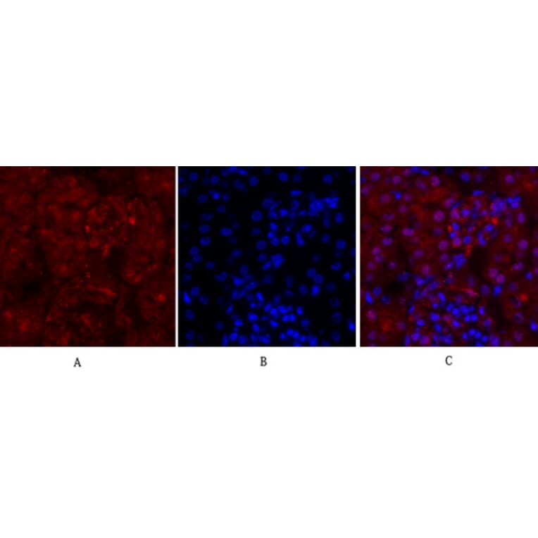
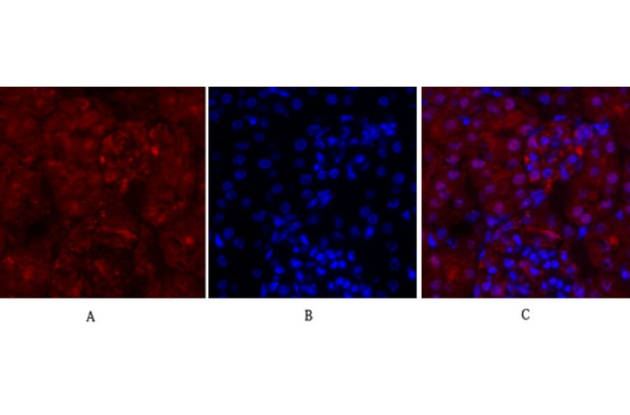
Immunofluorescence - Anti-Cyclin A Antibody (A94878)
Immunofluorescence analysis of mouse kidney tissue using Anti-Cyclin A Antibody (red) at 1:200 (4°C overnight). Cy3 labelled secondary antibody was used at 1:300 (RT 50min). Panel A: Target. Panel B: DAPI. Panel C: Merge.
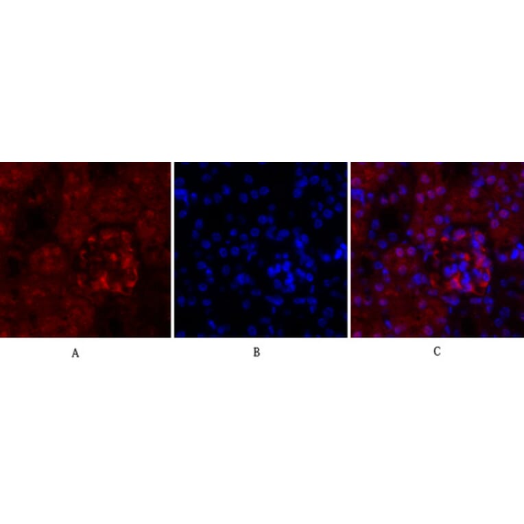
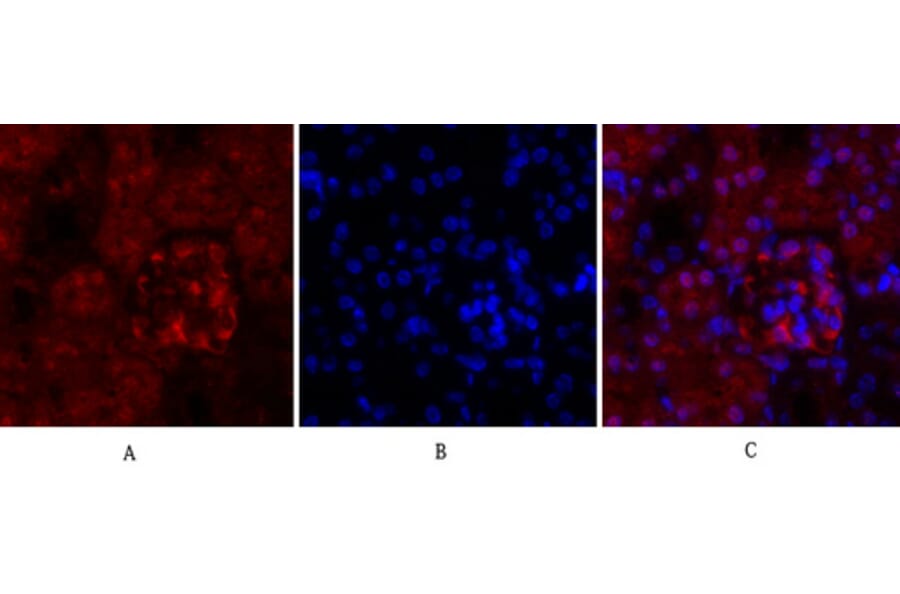
Immunofluorescence - Anti-Cyclin A Antibody (A94878)
Immunofluorescence analysis of mouse kidney tissue using Anti-Cyclin A Antibody (red) at 1:200 (4°C overnight). Cy3 labelled secondary antibody was used at 1:300 (RT 50min). Panel A: Target. Panel B: DAPI. Panel C: Merge.