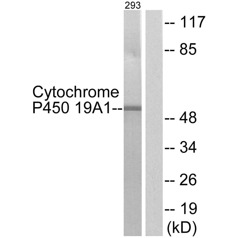
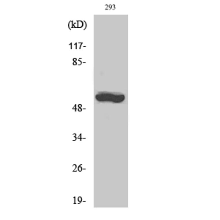
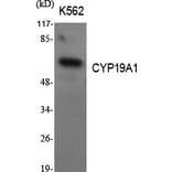
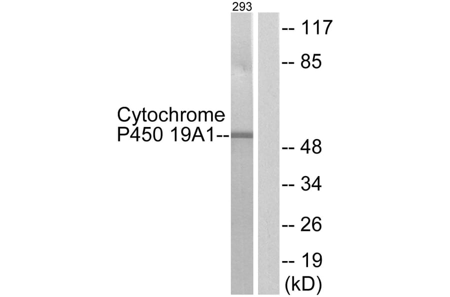
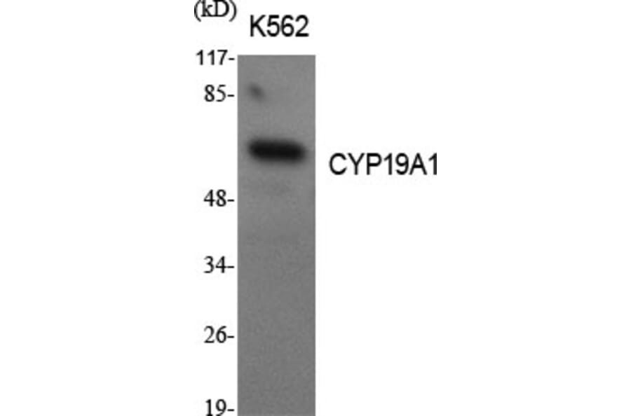
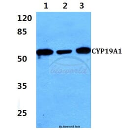
Western Blot - Anti-Cytochrome P450 19A1 Antibody (A93784)
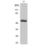
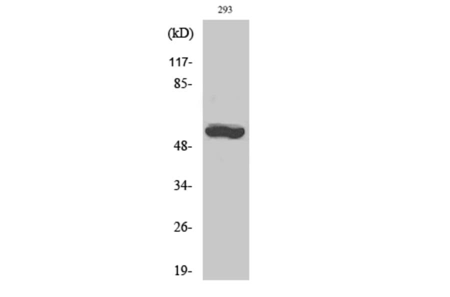
Western blot analysis of lysates from 293 cells using Anti-Cytochrome P450 19A1 Antibody. The right hand lane represents a negative control, where the antibody is blocked by the immunising peptide.
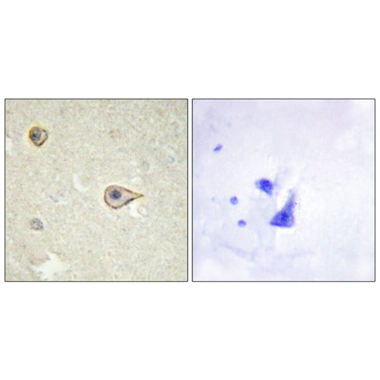
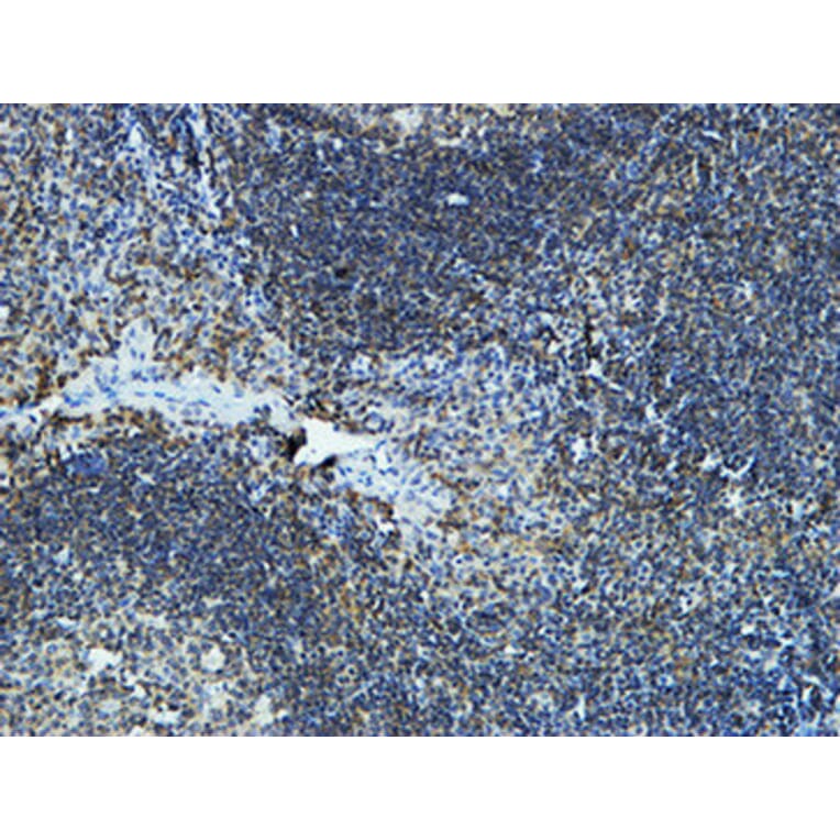
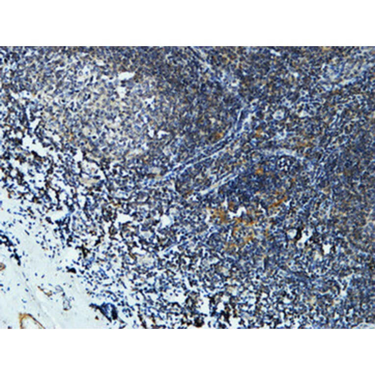
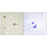
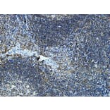
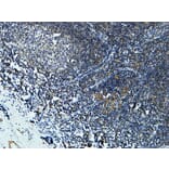
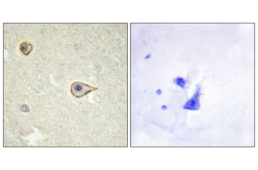
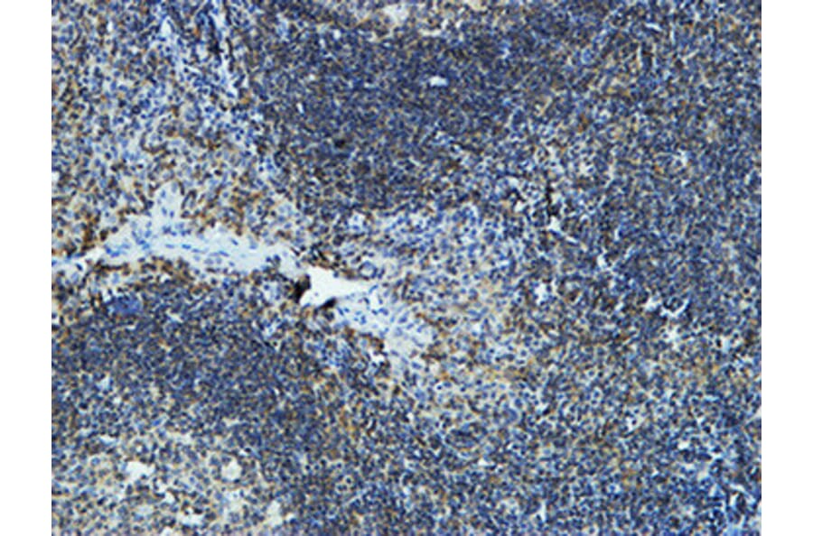
Immunohistochemical analysis of paraffin-embedded human brain tissue using Anti-Cytochrome P450 19A1 Antibody. The right hand panel represents a negative control, where the antibody was pre-incubated with the immunising peptide.
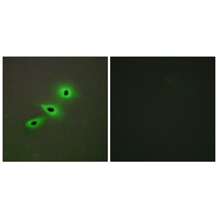
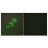
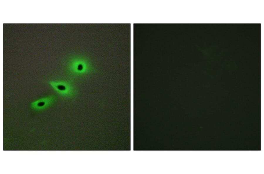
Immunofluorescence analysis of A549 cells using Anti-Cytochrome P450 19A1 Antibody. The right hand panel represents a negative control, where the antibody was pre-incubated with the immunising peptide.
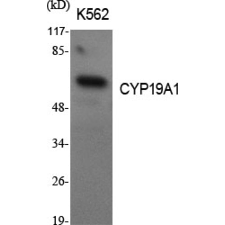
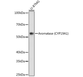
Western Blot - Anti-Cytochrome P450 19A1 Antibody (A93784)
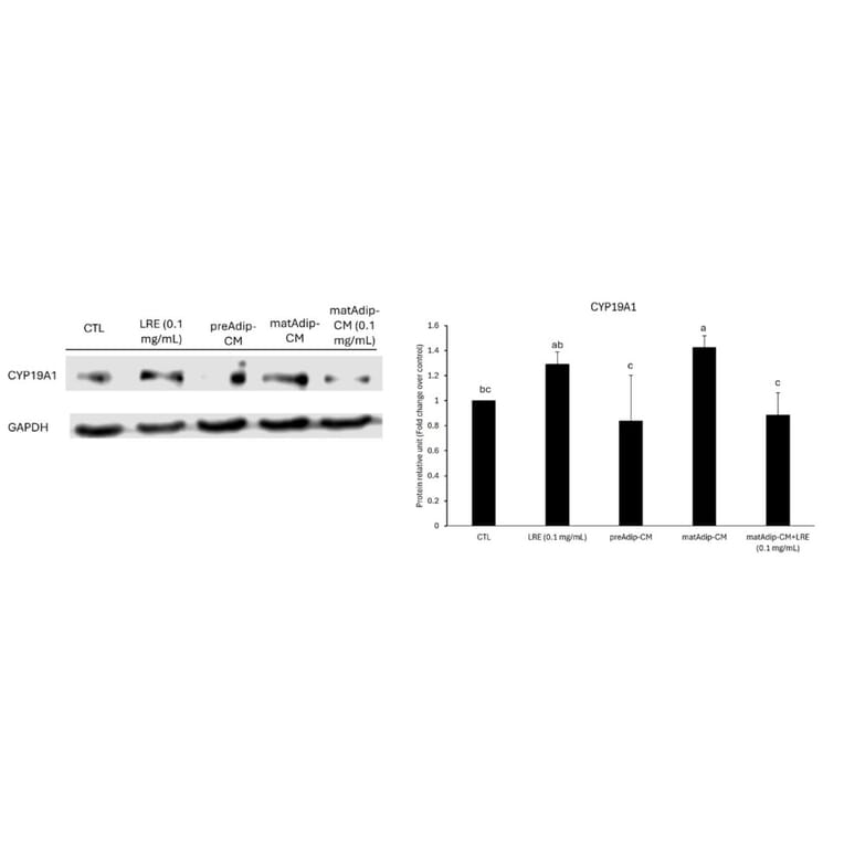
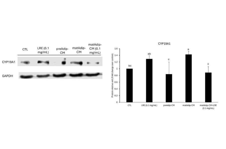
Protein expression of CYP19A1 in MCF-7 breast cancer cells under different treatment conditions. MCF-7 cells were treated for 48?h with LRE (0.1?mg/mL), conditioned medium from preadipocytes (preAdip-CM), mature adipocytes (matAdip-CM), or LRE-treated mature adipocytes (matAdip?+?LRE-CM), and compared with untreated control cells (CTL). CYP19A1 protein expression was assessed by Western blot, with GAPDH as loading control; the bar graph shows densitometric quantification. Values are expressed as mean?±?SD of three independent experiments (n?=?3). Different letters indicate statistically significant differences (p?
Figure 11B from Cianciosi et al. (2026). Originally published in Int J Food Sci Nutr, DOI: 10.1080/09637486.2026.2666404. Epub ahead of print.. Reproduced under CC BY 4.0.


![Western Blot - Anti-Aromatase Antibody [H4] (A280555) - Antibodies.com](https://cdn.antibodies.com/image/catalog/280/A280555_1.jpg?profile=product_alternative)

![SDS-PAGE - Anti-Aromatase Antibody [CYP19A1/4257] (A252297) - Antibodies.com](https://cdn.antibodies.com/image/catalog/248/A248315_1.jpg?profile=product_alternative)
![SDS-PAGE - Anti-Aromatase Antibody [CYP19A1/4257] - BSA and Azide free (A248315) - Antibodies.com](https://cdn.antibodies.com/image/catalog/251/A251497_1.jpg?profile=product_alternative)