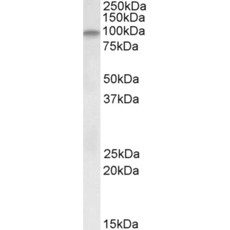
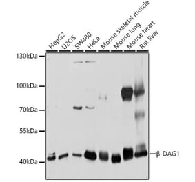
DAG1 expression in Human Skeletal Muscle lysate analyzed by western blot. Cells were lysed in RIPA buffer and 35µg protein was run per lane. Primary antibody incubation was performed with Anti-DAG1 Antibody (A82725) at 0.2µg/ml and detected by chemiluminescence.
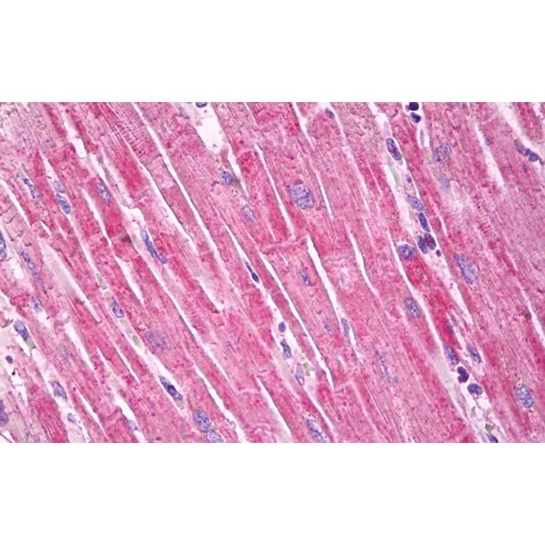
DAG1 expression in Human Heart analyzed by immunohistochemistry. Tissue was paraffin-embedded, and antigen retrieval was achieved by steaming in citrate buffer, pH 6. Staining was performed with Anti-DAG1 Antibody (A82725) at 5µg/ml and revealed with alkaline phosphatase (AP).
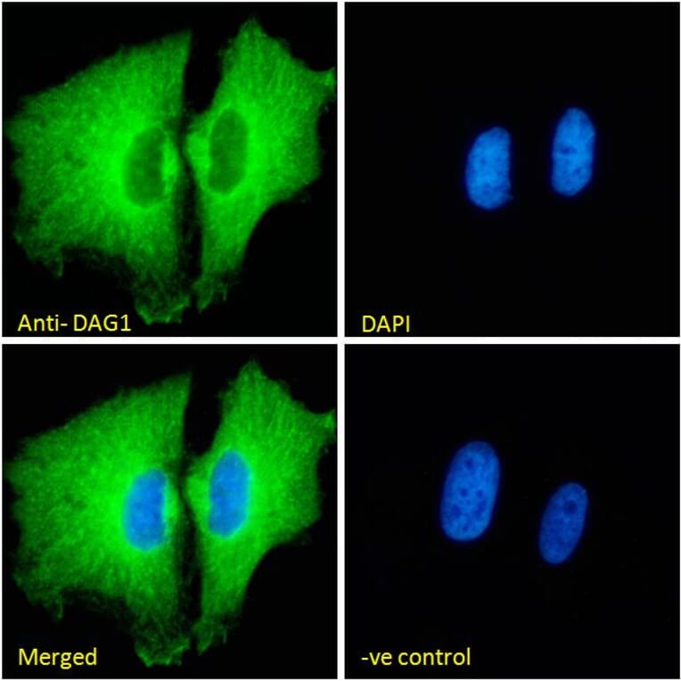
DAG1 expression in HeLa cells analyzed by immunofluorescence. Cells were permeabilized with 0.15% Triton. Staining was performed with Anti-DAG1 Antibody (A82725) at 10µg/ml for 1 hour and Alexa Fluor 488 secondary antibody at 2µg/ml. Cytoplasmic and ER staining shown and nuclei were stained with DAPI (blue). Negative control: Goat IgG Isotype Control at 10µg/ml followed by Alexa Fluor 488 secondary antibody at 2µg/ml.
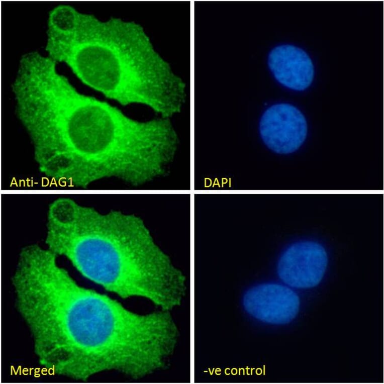
DAG1 expression in U2OS cells analyzed by immunofluorescence. Cells were permeabilized with 0.15% Triton. Staining was performed with Anti-DAG1 Antibody (A82725) at 10µg/ml for 1 hour and Alexa Fluor 488 secondary antibody at 2µg/ml. Cytoplasmic and ER staining shown and nuclei were stained with DAPI (blue). Negative control: Goat IgG Isotype Control at 10µg/ml followed by Alexa Fluor 488 secondary antibody at 2µg/ml.
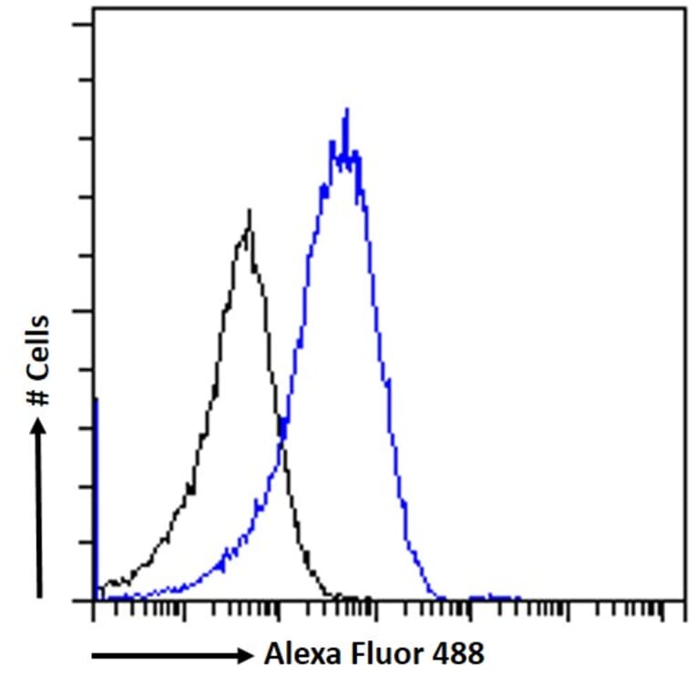
DAG1 expression in HeLa cells (blue line) analyzed by flow cytometry. Cells were fixed in PFA and permeabilized with 0.5% Triton. Staining was performed with Anti-DAG1 Antibody (A82725) at 10µg/ml for 1 hour and Alexa Fluor 488 secondary antibody at 1µg/ml. Negative Control: Goat IgG Isotype Control (black line) followed by Alexa Fluor 488 secondary antibody.
Recombinant human monoclonal antibody to Dystroglycan for use as a research grade Anti-Human Dystrog for ELISA, Flow Cytometry, Functional Studies and in vivo Research.
Recombinant human monoclonal antibody to Dystroglycan for use as a research grade Anti-Human Dystrog for ELISA, Flow Cytometry, Functional Studies and in vivo Research.


![SDS-PAGE - Anti-Dystroglycan Antibody [DAG-6F4] Biosimilar - BSA and Azide free (A339328) - Antibodies.com](https://cdn.antibodies.com/image/catalog/339/A339328_1.jpg?profile=product_alternative)
