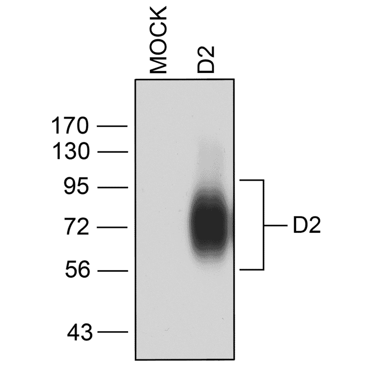
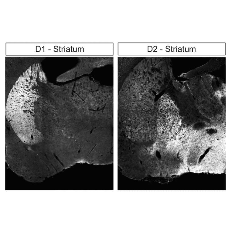
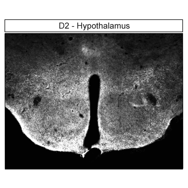
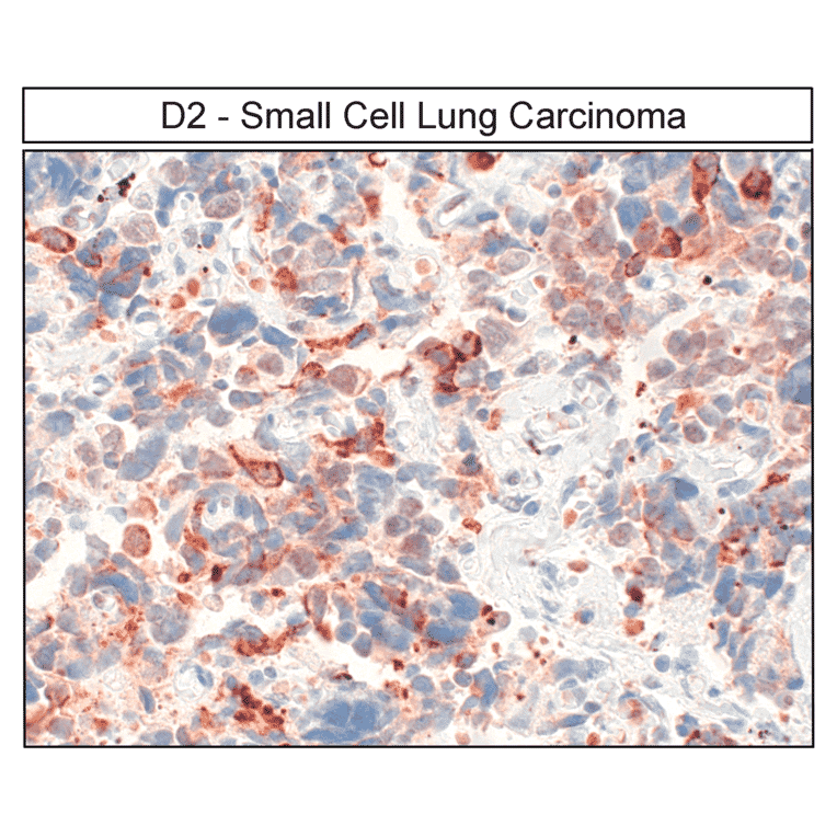
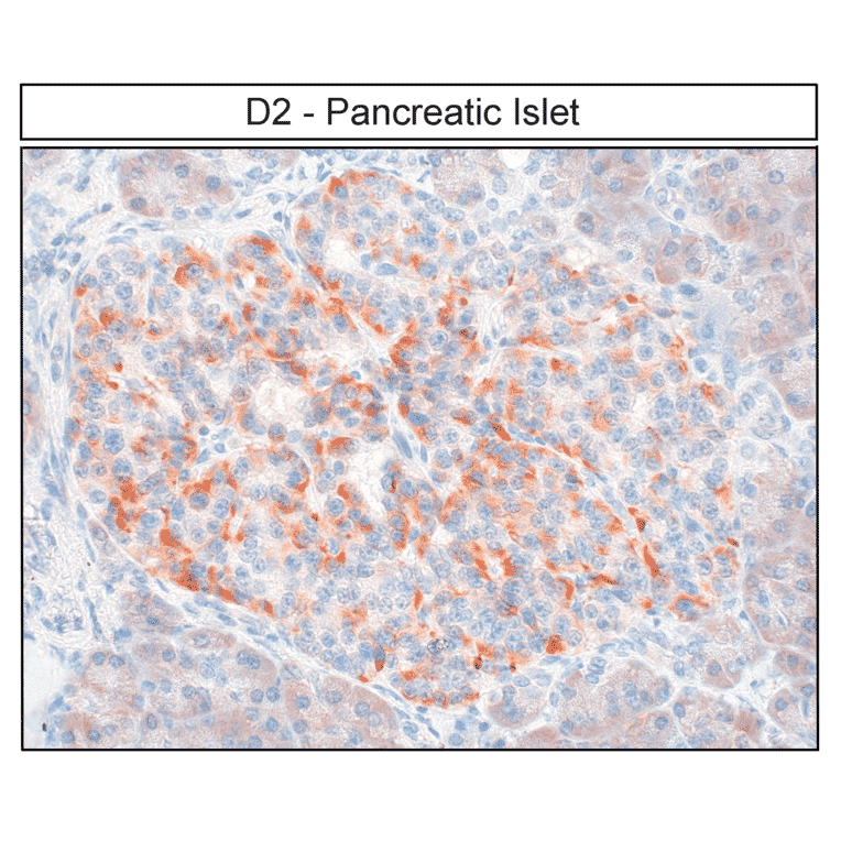
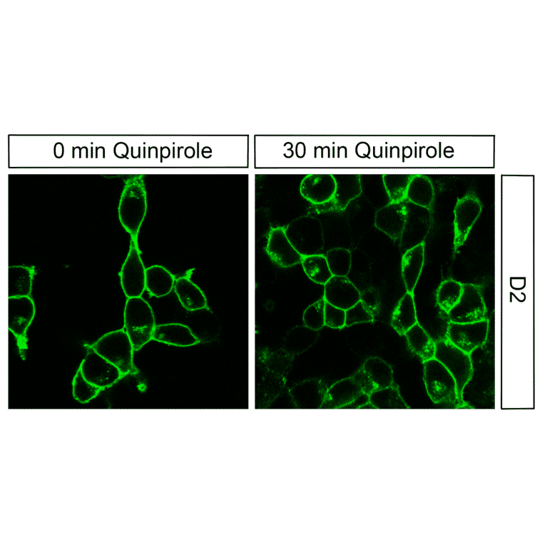
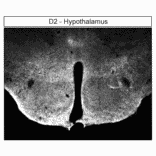
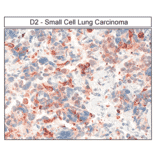
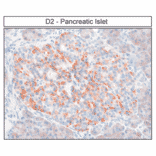
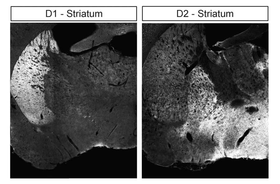
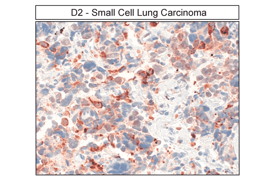
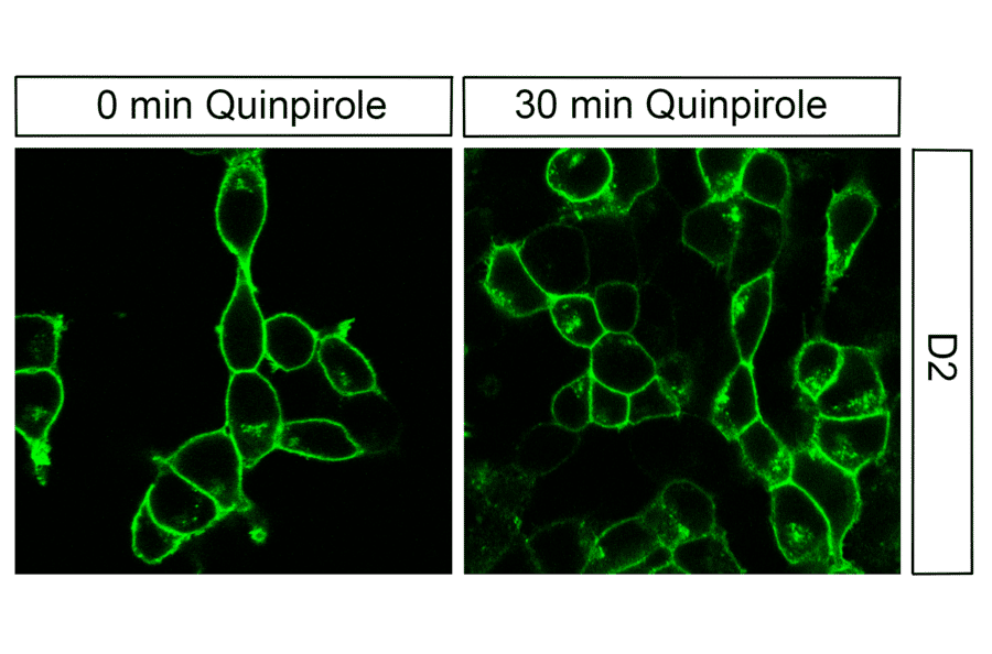
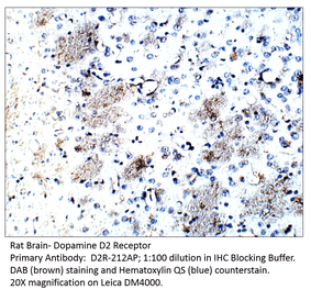
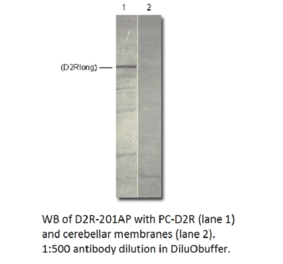
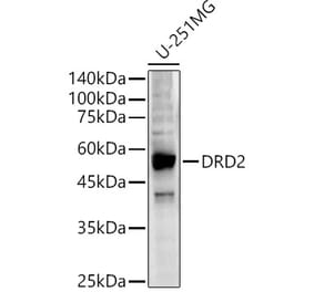
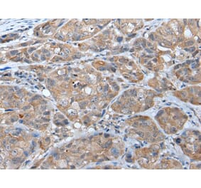
This antibody binds specifically to the third intracellular loop of human DRD2, detecting the non-phosphorylated isoform. It is validated for Western blot analysis, enables immunoprecipitation from brain tissue lysates, and supports immunohistochemistry in cultured cells and tissue sections.
Applications
WB, ICC, IHC
Dilutions
WB: 1,000, ICC: 1:200
Reactivity
Human, Mouse, Rat
Immunogen
Synthetic peptide corresponding to C-terminal of human DRD2. Immunogen range is 22-22 amino acids.
Host
Rabbit
Clonality
Polyclonal
Isotype
IgG
Conjugate
Unconjugated
Purification
Antigen affinity purification.
Concentration
Lot Specific
Product Form
Liquid
Formulation
Supplied in Dulbecco's PBS, pH 7.4, with 150 mM NaCl and 0.005% Sodium Azide.
Storage
Shipped at 4°C. Upon delivery aliquot and store at -20°C. Avoid freeze/thaw cycles.
Western blot confirmation of Dopamine Receptor 2 (DRD2) in transfected HEK293 cells. Lysates from native HEK293 cells (MOCK) or cells stably expressing DRD2 were immunoblotted with the phosphorylation-independent anti-DRD2 antibody (A334505) at a 1:1000 dilution.
Detection of Dopamine Receptor 1 (DRD1) and Dopamine Receptor 2 (DRD2) in striatum by immunohistochemistry. Striatal sections were dewaxed, microwaved in citric acid, and stained with either anti-DRD1 antibody (A334475) at 1:100 (left panel) or anti-DRD2 antibody (A334504) at 1:100 (right panel), followed by biotinylated anti-rabbit IgG and avidin-biotin solution. Color was developed with 3-amino-9-ethylcarbazole (AEC), and sections were counterstained with hematoxylin.
Immunohistochemical localization of Dopamine Receptor 2 (DRD2) in hypothalamus. Hypothalamic sections were dewaxed, microwaved in citric acid, and stained with anti-DRD2 antibody (A334505-I) at 1:100, followed by biotinylated anti-rabbit IgG and avidin-biotin solution. Color was developed with 3-amino-9-ethylcarbazole (AEC), and sections were counterstained with hematoxylin. DRD2 receptors were detected in a distinct population of neuroendocrine tumor cells.
Detection of Dopamine Receptor 2 (DRD2) in human small cell lung carcinoma by immunohistochemistry. Tumor sections were dewaxed, microwaved in citric acid, and incubated with anti-DRD2 antibody (A334505) at 1:100, followed by biotinylated anti-rabbit IgG and avidin-biotin solution. Color was developed with 3-amino-9-ethylcarbazole (AEC), and sections were counterstained with hematoxylin. DRD2 receptors were observed in a specific subset of carcinoma cells.
Immunohistochemical localization of Dopamine Receptor 2 (DRD2) in human pancreatic islets. Pancreatic sections were dewaxed, microwaved in citric acid, and stained with anti-DRD2 antibody (A334505) at 1:100, followed by biotinylated anti-rabbit IgG and avidin-biotin solution. Color was developed with 3-amino-9-ethylcarbazole (AEC), and sections were counterstained with hematoxylin. DRD2 receptors were detected in pancreatic islet cells.
Immunocytochemical analysis of Dopamine Receptor 2 (DRD2) in transfected HEK293 cells. HEK293 cells stably expressing DRD2 were untreated or treated with 10 µM Quinpirole for 30 min and stained with anti-DRD2 antibody (A334505) at 1:200. DRD2 receptors were localized to the plasma membrane in untreated cells (0 min) and in both the plasma membrane and perinuclear vesicular clusters after 30 min of Quinpirole exposure.