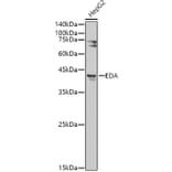
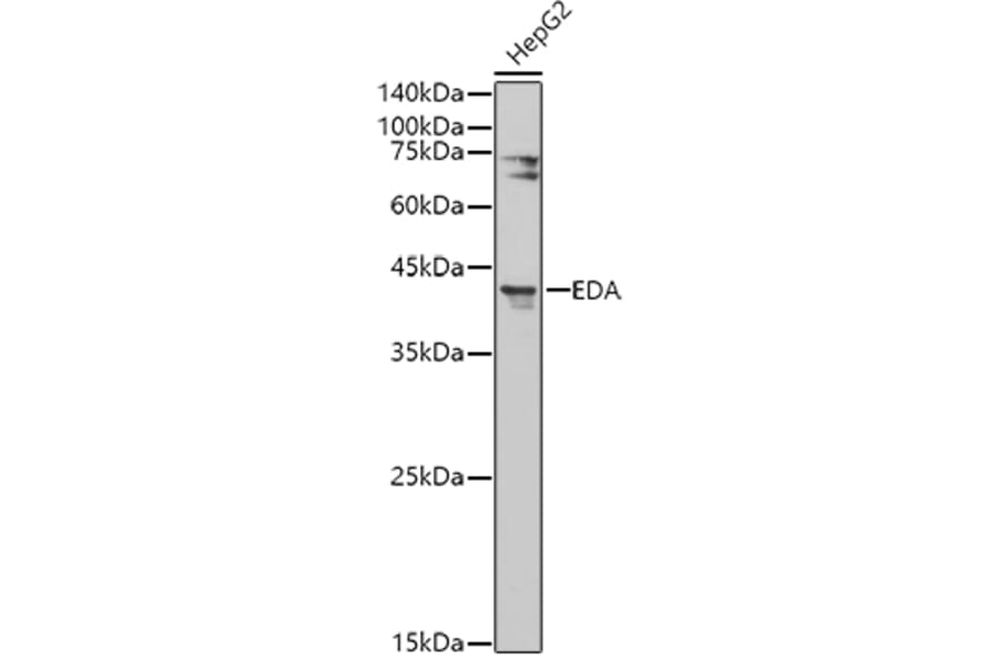
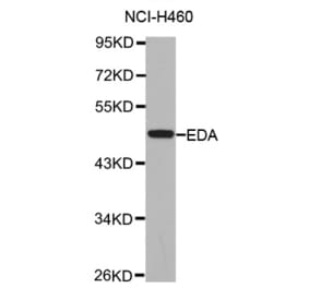
Western blot analysis of extracts of HepG2 cells, using Anti-EDA Antibody (A14246) at 1:500 dilution. The secondary antibody was Goat Anti-Rabbit IgG H&L Antibody (HRP) at 1:10,000 dilution. Lysates/proteins were present at 25µg per lane. The blocking buffer used was 3% non-fat dry milk in TBST. Detection was with a ECL Basic Kit. Exposure time: 180s.
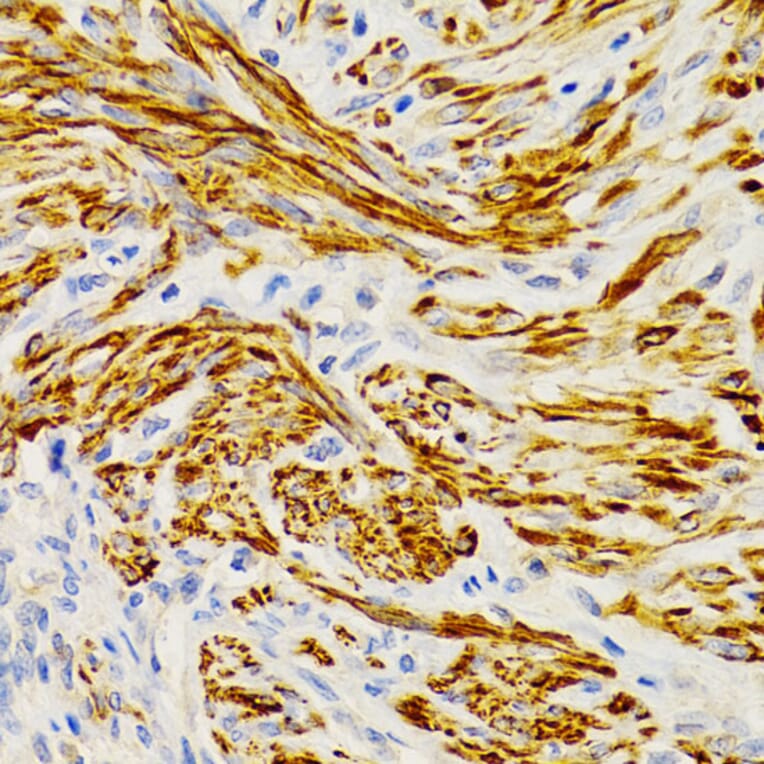
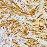
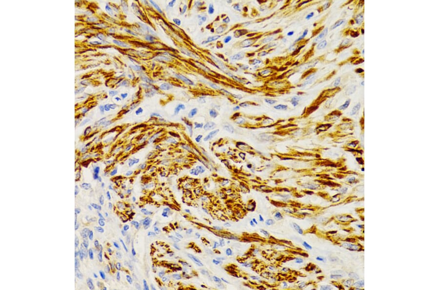
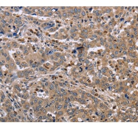
Immunohistochemistry analysis of paraffin-embedded human uterine cancer using Anti-EDA Antibody (A14246) at a dilution of 1:200 (40x lens). Perform microwave antigen retrieval with 10 mM PBS buffer pH 7.2 before commencing with IHC staining protocol.
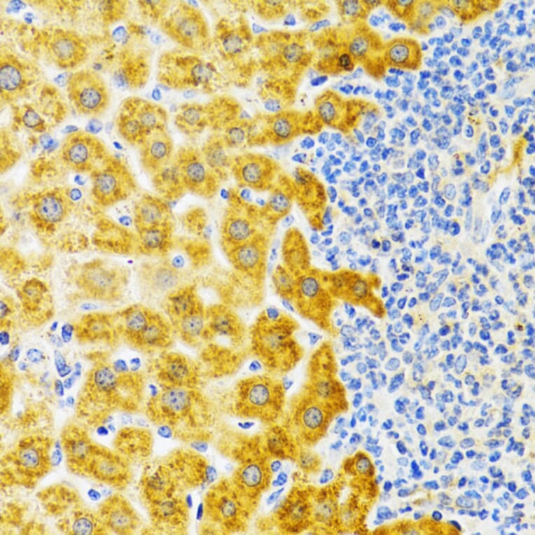
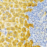
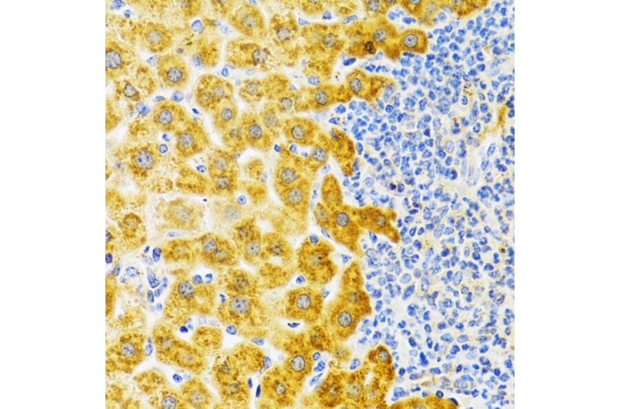
Immunohistochemistry analysis of paraffin-embedded rat liver using Anti-EDA Antibody (A14246) at a dilution of 1:100 (40x lens). Perform microwave antigen retrieval with 10 mM PBS buffer pH 7.2 before commencing with IHC staining protocol.
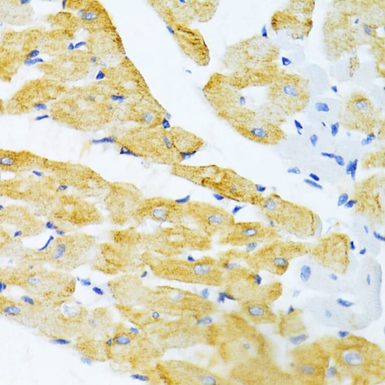
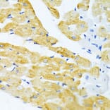
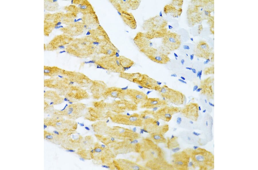
Immunohistochemistry analysis of paraffin-embedded mouse heart using Anti-EDA Antibody (A14246) at a dilution of 1:100 (40x lens). Perform microwave antigen retrieval with 10 mM PBS buffer pH 7.2 before commencing with IHC staining protocol.
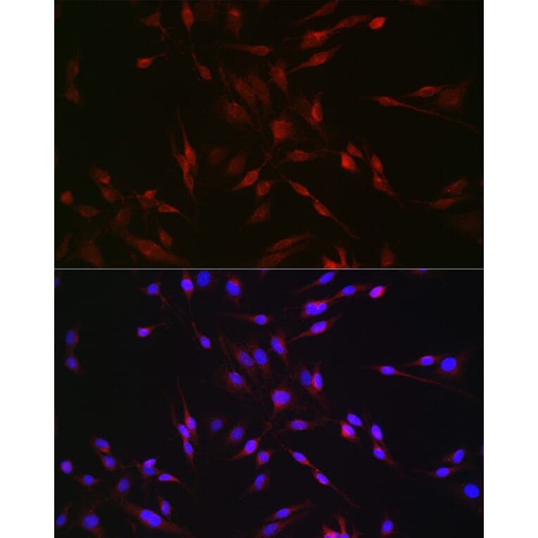
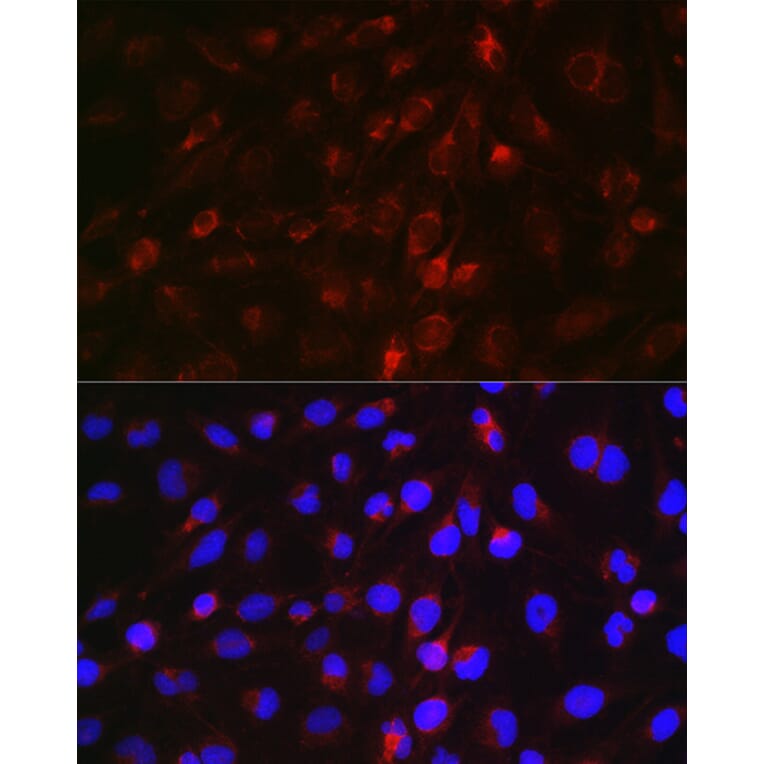
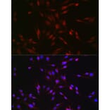
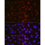
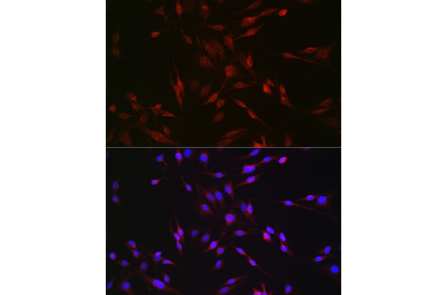
Immunofluorescence analysis of C6 cells using Anti-EDA Antibody (A14246) at a dilution of 1:50 (40x lens). DAPI was used to stain the cell nuclei (blue).
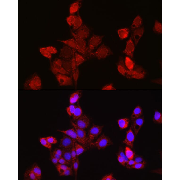
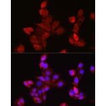
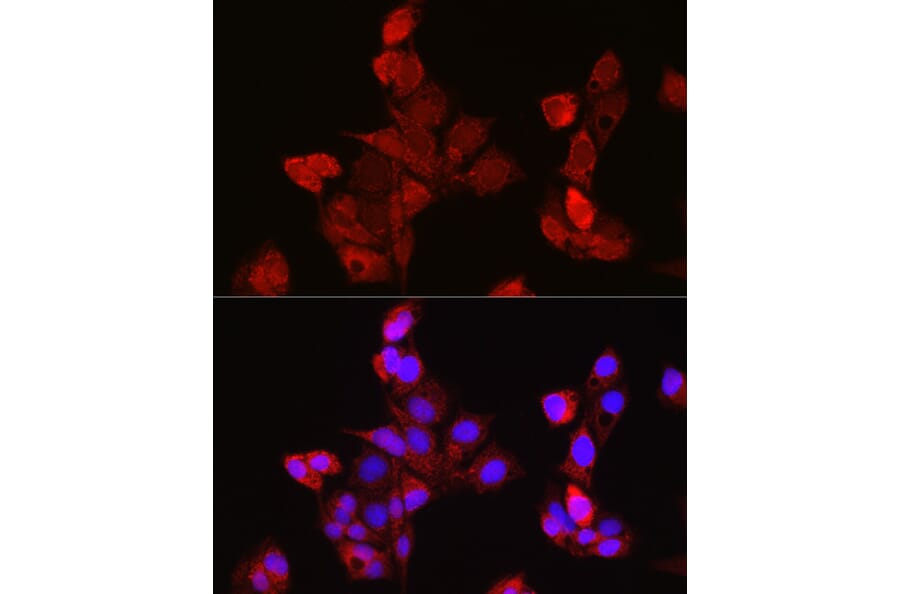
Immunofluorescence analysis of HepG2 cells using Anti-EDA Antibody (A14246) at a dilution of 1:50 (40x lens). DAPI was used to stain the cell nuclei (blue).
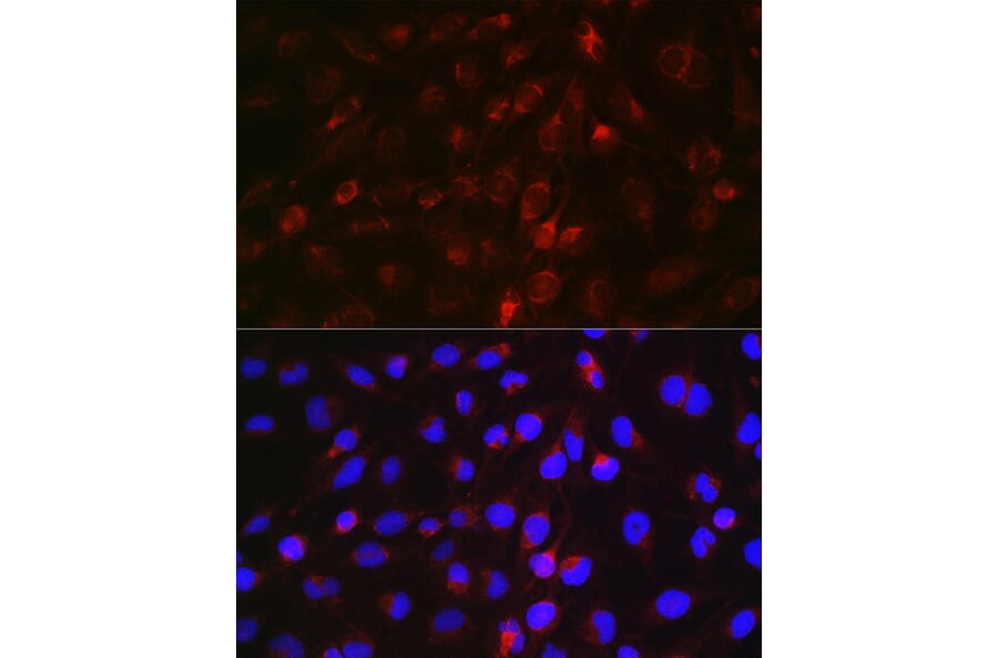
Immunofluorescence analysis of U-251MG cells using Anti-EDA Antibody (A14246) at a dilution of 1:50 (40x lens). DAPI was used to stain the cell nuclei (blue).


![Flow Cytometry - Anti-EDA Chimeric Antibody [DMC388] - Azide free (A318760) - Antibodies.com](https://cdn.antibodies.com/image/catalog/318/A318760_1.jpg?profile=product_alternative)