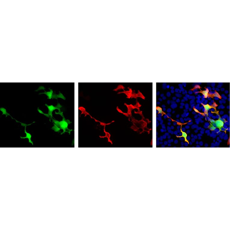
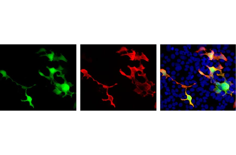
Transfected HEK293 cells which overexpress EosFP protein were stained with Anti-EosFP Antibody and viewed in a fluorescence microscope. Cells which are transfected with EosFP (left panel) are bright green. On staining with Anti-EosFP Antibody in red (middle panel), EosFP expressing cells appear orange (right panel). Most HEK293 cells are not transfected so only the nucleus of these cells can be visualized with a blue DNA stain.
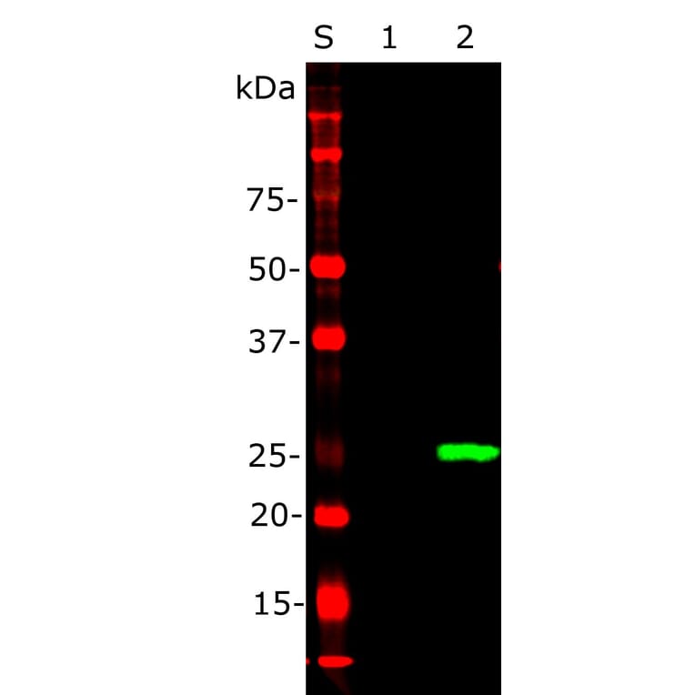
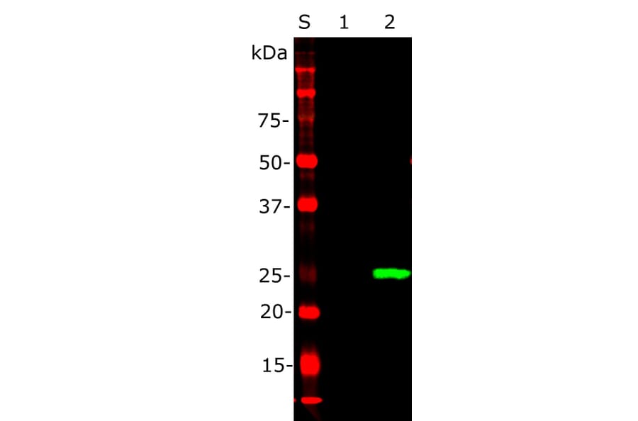
Western blot analysis of Anti-EosFP Antibody: 1: Non-transfected HEK293 crude homogenate. 2: Crude homogenate of transfected HEK293 cells which overexpress protein EosFP. S: Protein standard of indicated molecular weight. Anti-EosFP Antibody, at a dilution of 1: 1,000) reveals a strong clean band at ~25 kDa corresponding to EosFP in transfected cells, which is absent from non-transfected cells.
Publishing research using Anti-EosFP Antibody (A85295)? Please let us know so that we can list the citation on this page.