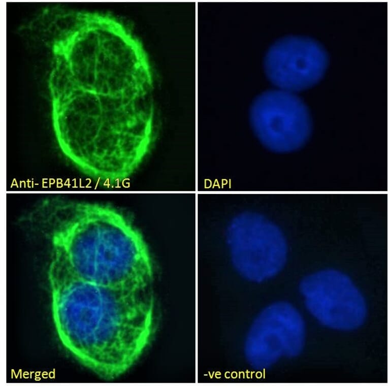
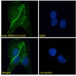
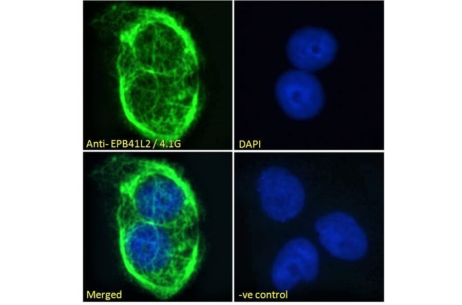
EPB41L2 expression in A431 cells analyzed by immunofluorescence. Cells were permeabilized with 0.15% Triton. Staining was performed with Anti-EPB41L2 Antibody (A84421) at 10µg/ml for 1 hour and Alexa Fluor 488 secondary antibody at 2µg/ml. Plasma membrane staining shown and nuclei were stained with DAPI (blue). Negative control: Goat IgG Isotype Control at 10µg/ml followed by Alexa Fluor 488 secondary antibody at 2µg/ml.
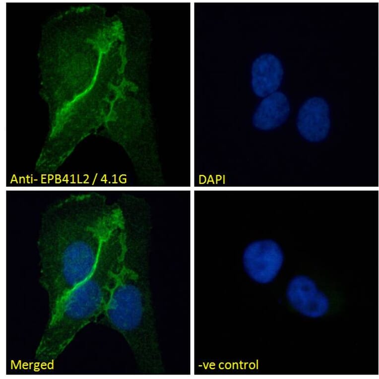
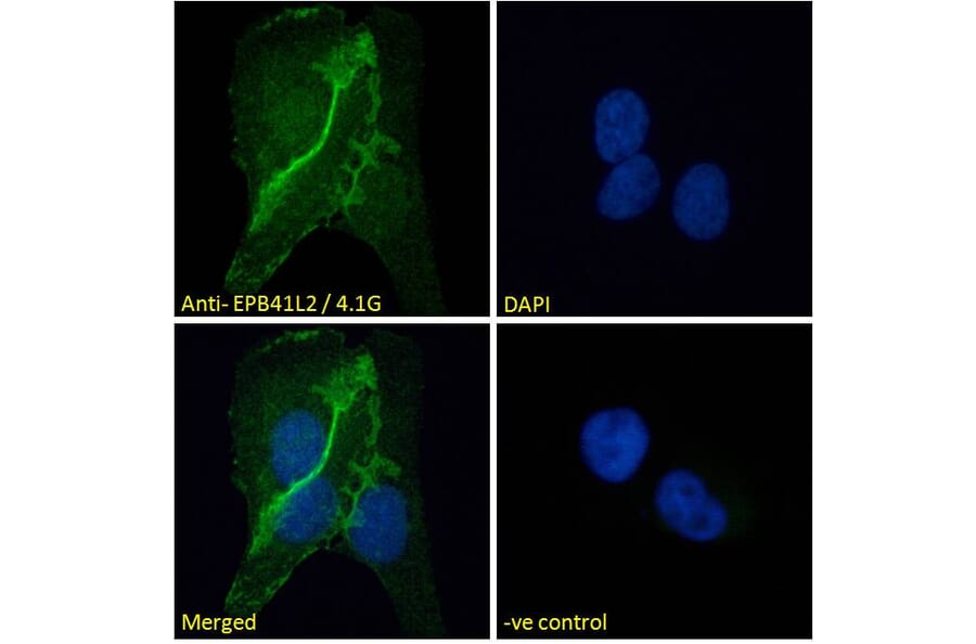
EPB41L2 expression in U2OS cells analyzed by immunofluorescence. Cells were permeabilized with 0.15% Triton. Staining was performed with Anti-EPB41L2 Antibody (A84421) at 10µg/ml for 1 hour and Alexa Fluor 488 secondary antibody at 2µg/ml. Cell junction staining shown and nuclei were stained with DAPI (blue). Negative control: Goat IgG Isotype Control at 10µg/ml followed by Alexa Fluor 488 secondary antibody at 2µg/ml.
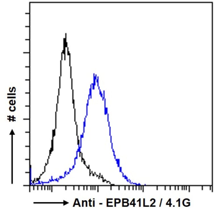
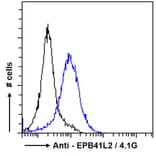
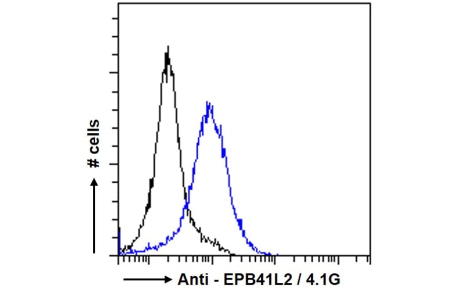
EPB41L2 expression in A431 cells (blue line) analyzed by flow cytometry. Cells were fixed in PFA and permeabilized with 0.5% Triton. Staining was performed with Anti-EPB41L2 Antibody (A84421) at 10µg/ml for 1 hour and Alexa Fluor 488 secondary antibody at 1µg/ml. Negative Control: Goat IgG Isotype Control (black line) followed by Alexa Fluor 488 secondary antibody.
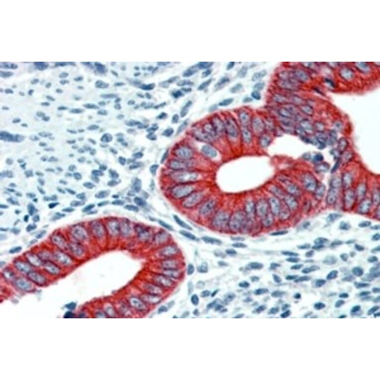
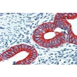
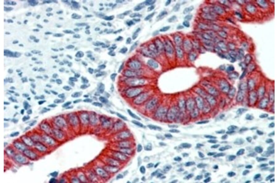
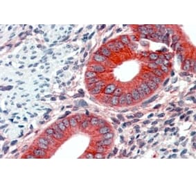
EPB41L2 expression in Human Uterus analyzed by immunohistochemistry. Tissue was paraffin-embedded, and antigen retrieval was achieved by steaming in citrate buffer, pH 6. Staining was performed with Anti-EPB41L2 Antibody (A84421) at 2.5µg/ml and revealed with alkaline phosphatase (AP).
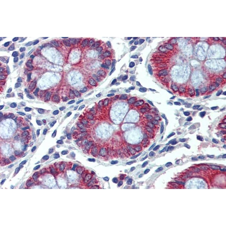
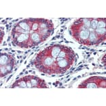
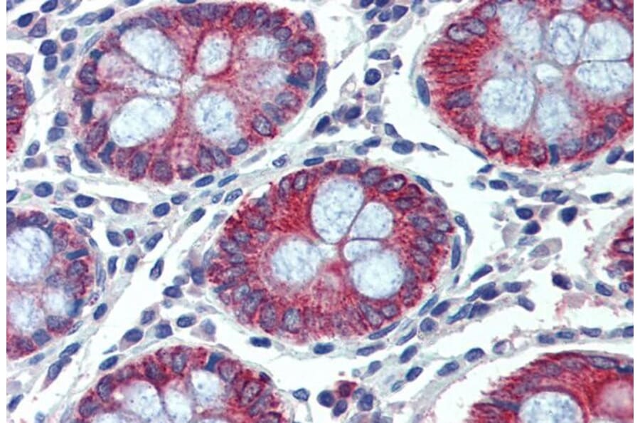
EPB41L2 expression in Human Colon analyzed by immunohistochemistry. Tissue was paraffin-embedded, and antigen retrieval was achieved by steaming in citrate buffer, pH 6. Staining was performed with Anti-EPB41L2 Antibody (A84421) at 2.5µg/ml and revealed with alkaline phosphatase (AP).