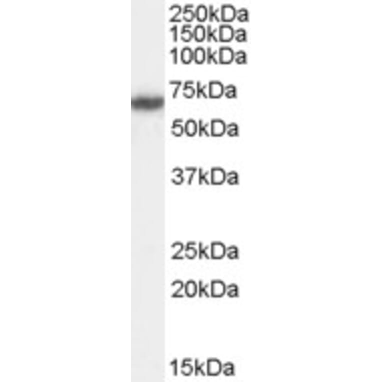
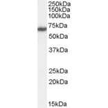
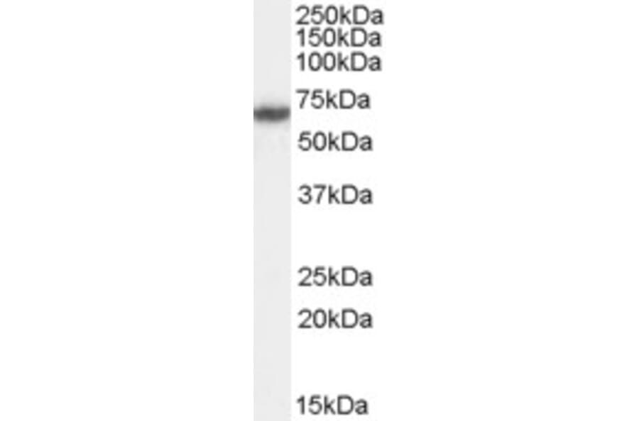
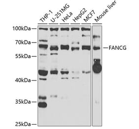
FANCG expression in HeLa cell lysate analyzed by western blot. Cells were lysed in RIPA buffer and 35µg protein was run per lane. Primary antibody incubation was performed for 1 hour with Anti-FANCG Antibody (A83967) at 0.5µg/ml and detected by chemiluminescence.
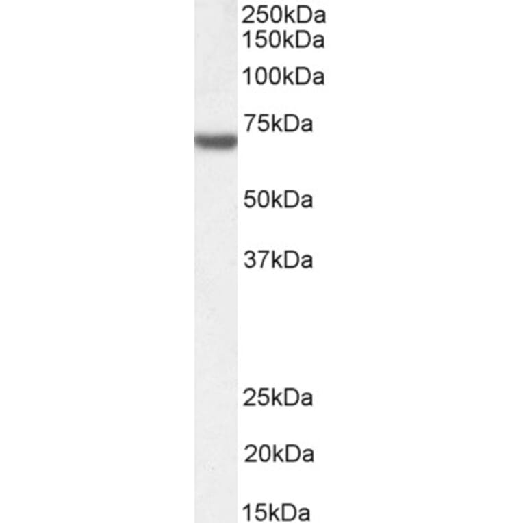
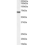
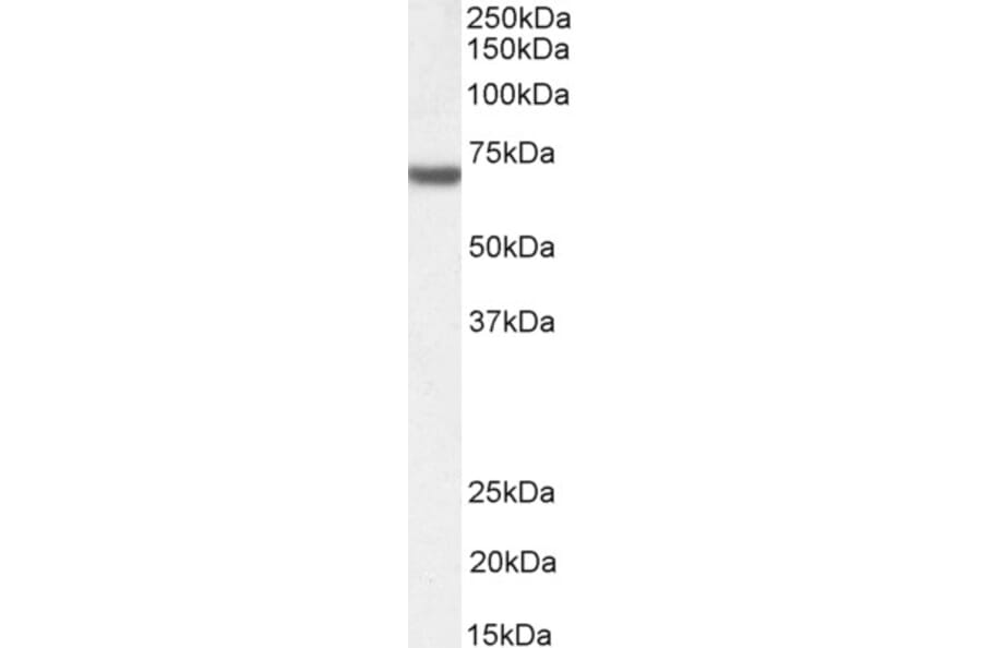
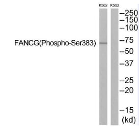
FANCG expression in Jurkat nuclear cell lysate analyzed by western blot. Cells were lysed in RIPA buffer and 35µg protein was run per lane. Primary antibody incubation was performed for 1 hour with Anti-FANCG Antibody (A83967) at 1µg/ml and detected by chemiluminescence.
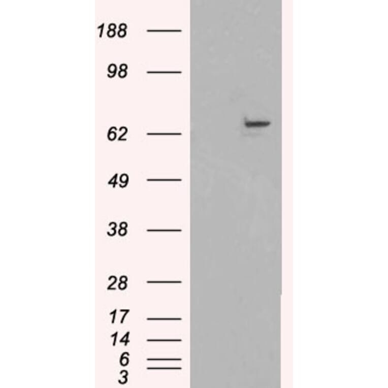
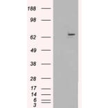
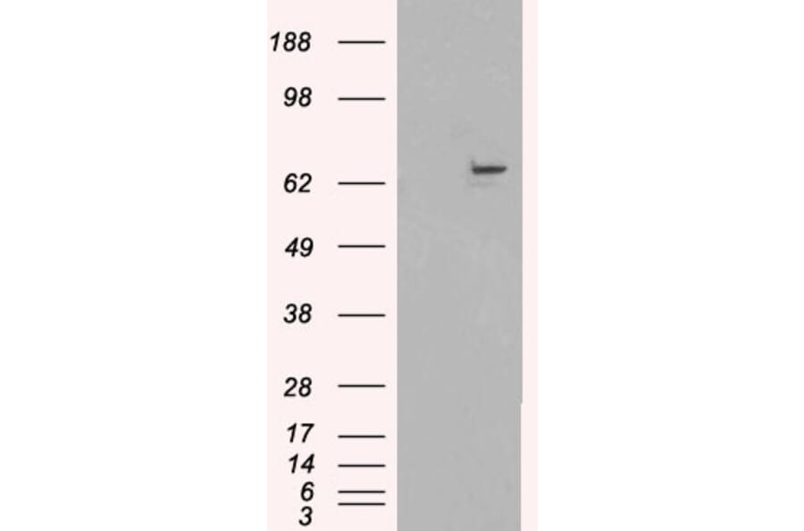
FANCG expression in HEK293 cell lysate analyzed by western blot. Cells were transfected to overexpress FANCG and incubated with Anti-FANCG Antibody (A83967). Control: Mock-transfected HEK293 cell lysate incubated with Anti-FANCG Antibody (A83967) (first lane).
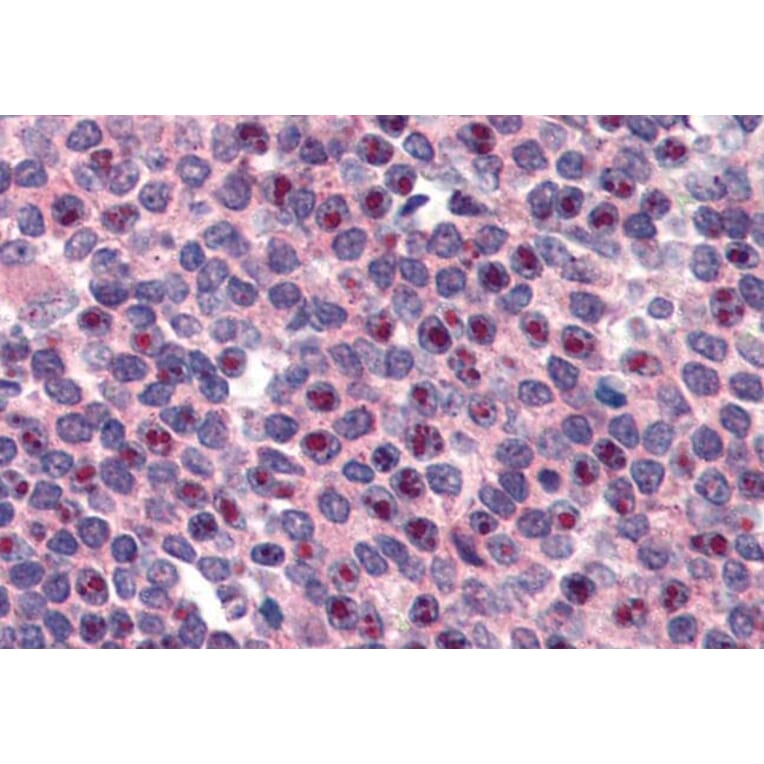
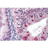
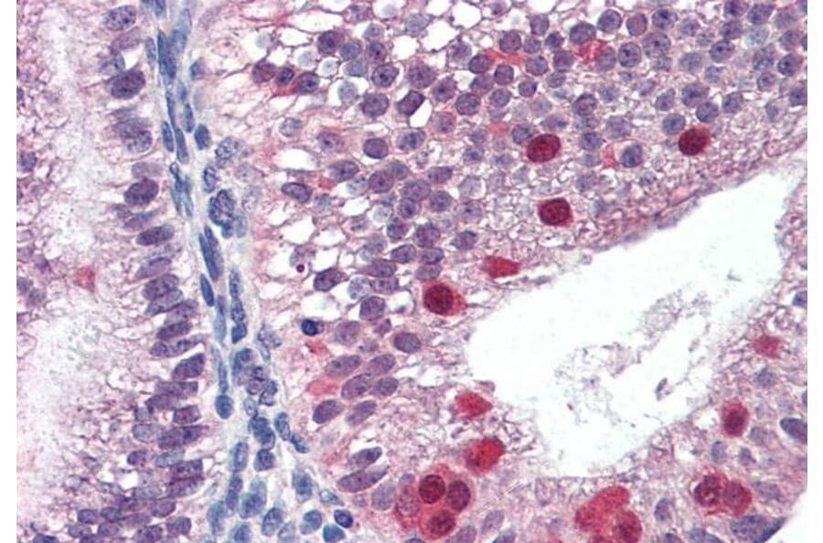
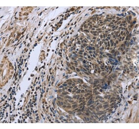
FANCG expression in Human Uterus analyzed by immunohistochemistry. Tissue was paraffin-embedded, and antigen retrieval was achieved by steaming in citrate buffer, pH 6. Staining was performed with Anti-FANCG Antibody (A83967) at 3µg/ml and revealed with alkaline phosphatase (AP).
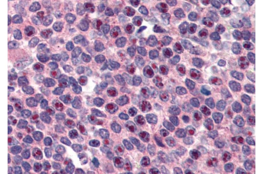
FANCG expression in Human Spleen analyzed by immunohistochemistry. Tissue was paraffin-embedded, and antigen retrieval was achieved by steaming in citrate buffer, pH 6. Staining was performed with Anti-FANCG Antibody (A83967) at 3µg/ml and revealed with alkaline phosphatase (AP).