This polyclonal rabbit anti-FLAG antibody was raised against the FLAG epitope tag and specifically recognizes the DYKDDDDK sequence fused to the N- or C-terminus of recombinant proteins.
CF® Dye and biotin conjugates: Add 0.5 ml dH2O for reconstitution. HRP or DNP conjugates: Add 1 ml dH2O for reconstitution.
Formulation
Supplied in Phosphate Buffered Saline containing 50% glycerol, 2 mg/ml BSA, and 0.05% Sodium Azide.
Storage
Shipped at +4°C. Upon delivery aliquot and store at -20°C. Avoid freeze/thaw cycles. This product is also photosensitive and should be protected from light. Should this product contain a precipitate we recommend microcentrifugation before use. CF® Dyes are guaranteed for at least 6 months from data of receipt when stored correctly.
General Notes
Looking for a specific protein conjugate to simplify your workflow? We offer a library of over 2,000 targets conjugated to your choice of CF® dye. To enquire about a custom product, contact us directly.
Synonyms
DDDDK epitope tag, DDDDK tag, DDDK, ddk, DYKDDDDK, DYKDDDDK epitope tag, DYKDDDDK tag, ECS epitope tag, ECS tag, Enterokinase Cleavage Site epitope tag, Enterokinase Cleavage Site tag, FLAG
Disclaimer
This product is for research use only. It is not intended for diagnostic or therapeutic use.
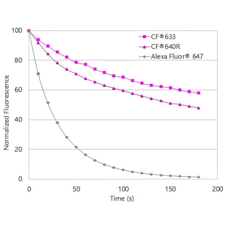
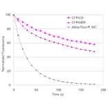
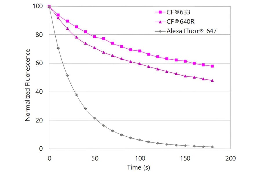
CF®640R exhibits substantially greater photostability than Alexa Fluor® 647. Jurkat cells were stained with mouse anti-CD3 primary antibody followed by goat anti-mouse secondary antibodies conjugated to the indicated dyes. Cells were continuously illuminated using a mercury arc lamp with a Cy®5 filter cube, with images acquired every 15 seconds for 5 minutes. Fluorescence intensity was normalized to the initial time point.
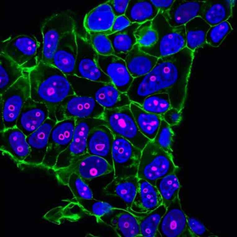
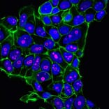
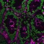
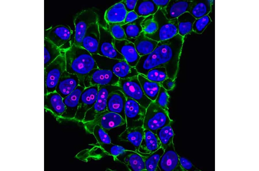
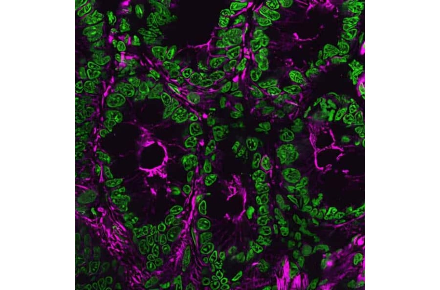
MCF-7 cells stained with CF®640R-conjugated anti-Cyclin B1 antibody to label nuclei and nucleoli (magenta), CF®488A phalloidin to visualize actin filaments (green), and Hoechst to stain DNA (blue).
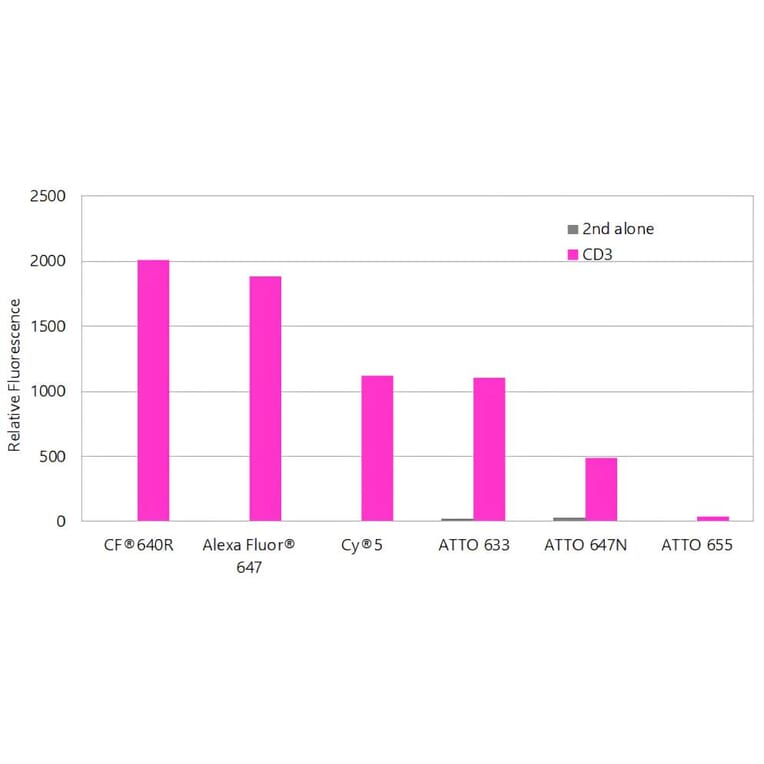
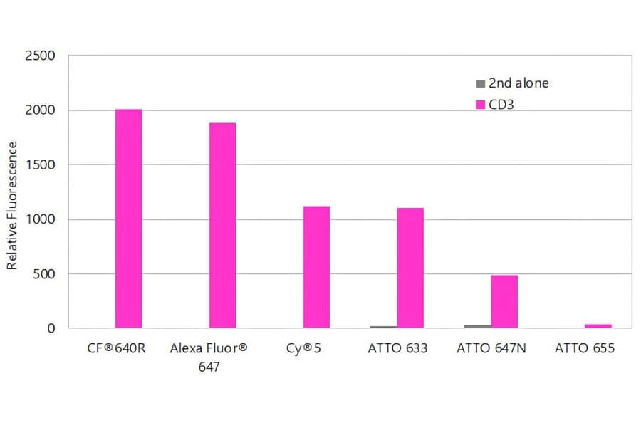
CF®640R produces brighter antibody conjugates than other far-red dyes. Jurkat cells were stained with mouse anti-CD3 primary antibody or without primary antibody as a control, followed by goat anti-mouse secondary antibodies conjugated to the indicated dyes. Fluorescence was analyzed on a BD FACSCalibur™ flow cytometer using the FL4 detection channel, and bars represent geometric mean fluorescence intensity.
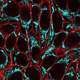
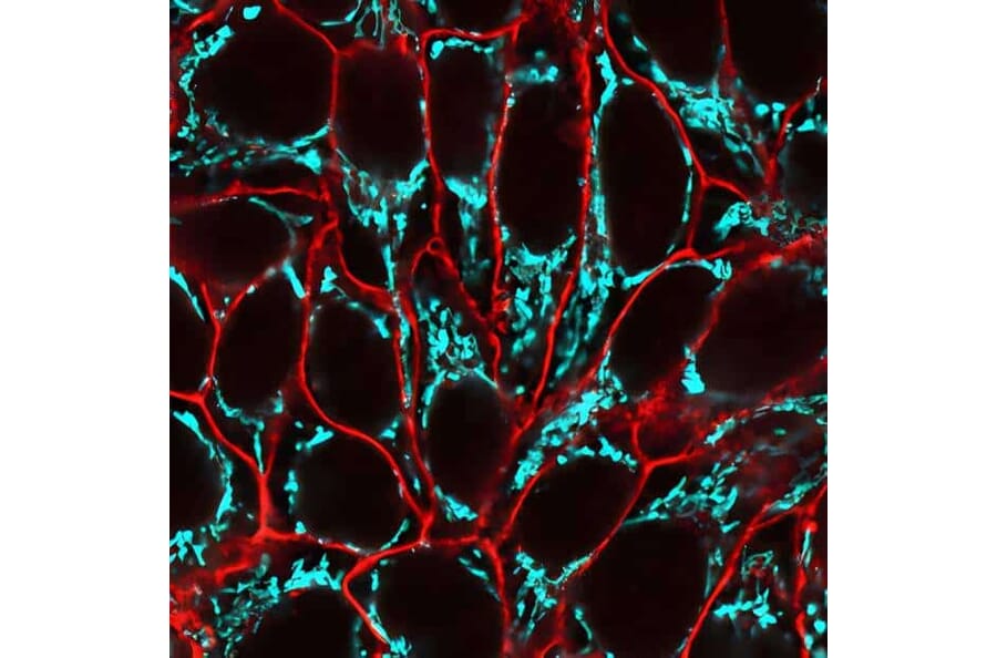
HeLa cells labeled with CellBrite® Fix 555 to stain the plasma membrane (red), fixed, and subsequently stained with CF®640R–conjugated anti-mitochondrial marker antibody (clone 113-1) to label mitochondria (cyan).
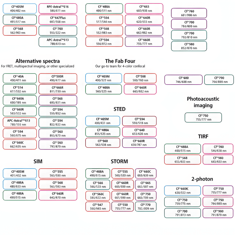
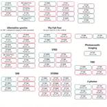
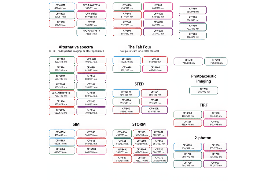
This chart summarizes commonly used CF® dyes grouped by their suitability for specific imaging modalities, including alternative spectra applications, four-color confocal imaging, near-infrared western blotting, photoacoustic imaging, STED, SIM, STORM, TIRF, and two-photon microscopy. Each dye is shown with its characteristic excitation and emission wavelengths (nm), providing a practical reference for selecting spectrally compatible dyes and optimizing multicolor experimental design across a range of fluorescence techniques.
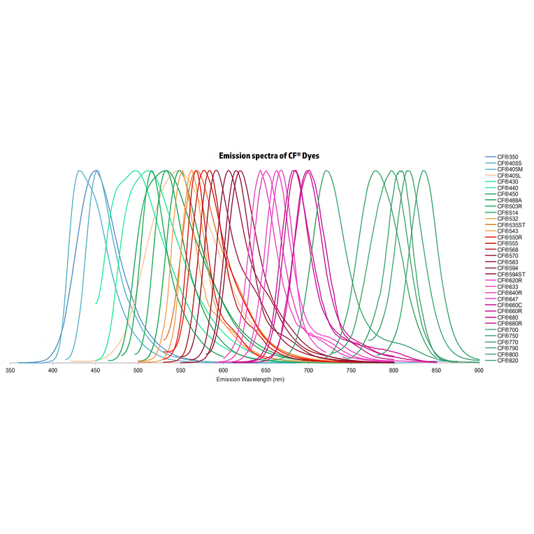
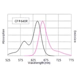
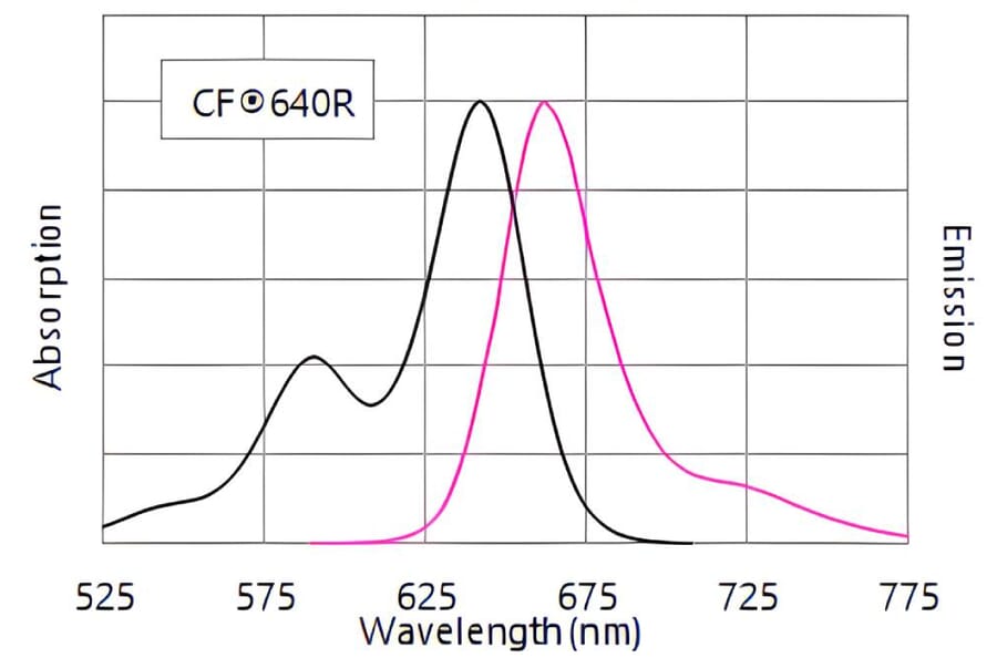
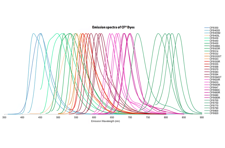
Normalized emission spectra of the CF® dye family spanning the visible to near-infrared range are shown, illustrating the spectral diversity and overlap between dyes. Curves represent relative fluorescence intensity as a function of emission wavelength (nm), with peak positions corresponding to each dye’s characteristic emission maximum. This reference highlights the broad coverage of CF® dyes for multicolor fluorescence applications and aids in selecting compatible dye combinations for imaging, flow cytometry, and other fluorescence-based assays.
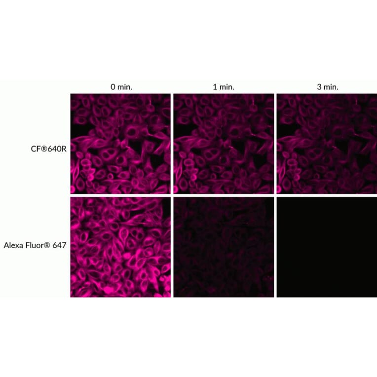
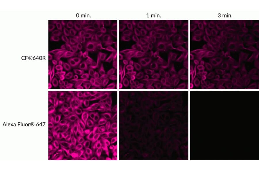
Relative photostability of CF®640R and Alexa Fluor® 647 fluorescence. HeLa cells were stained with mouse anti-tubulin primary antibody followed by goat anti-mouse secondary antibodies conjugated to the indicated dyes. Cells were continuously illuminated using a mercury arc lamp with a Cy®5 filter set, and images were acquired at time zero and after 1 and 3 minutes of light exposure.