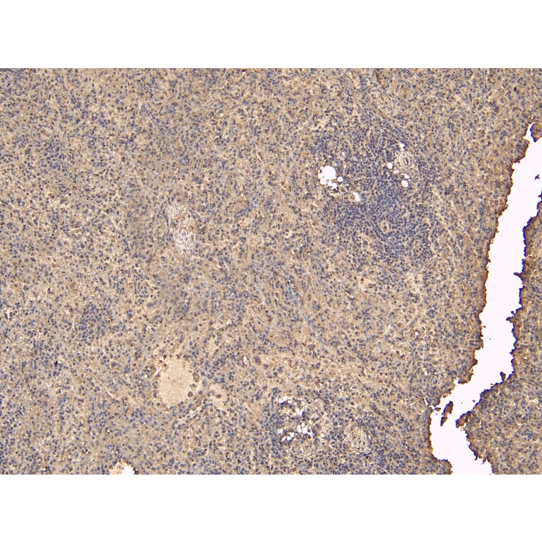
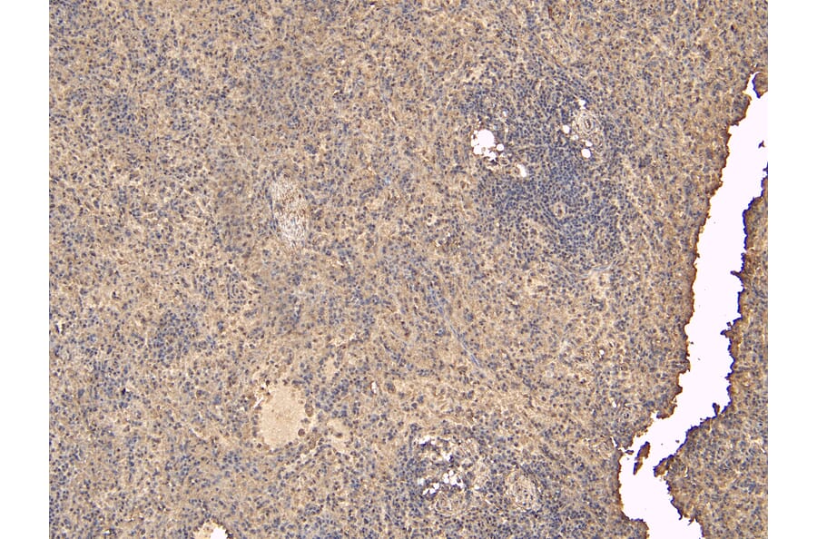
FLAP expression in Human Spleen analyzed by immunohistochemistry. Tissue was paraffin-embedded, and antigen retrieval was achieved by heating in citrate buffer, pH 6. Staining was performed with Anti-FLAP Antibody (A84031) at 6µg/ml and revealed with horseradish peroxidase (HRP).
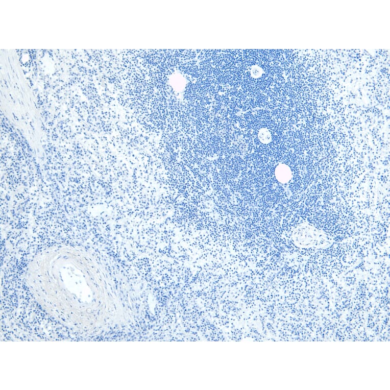
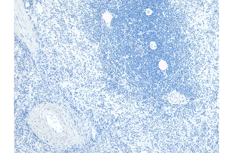
Negative control for FLAP expression in Human Spleen analyzed by immunohistochemistry. Tissue was paraffin-embedded, and staining procedure was performed in the absence of primary antibody.
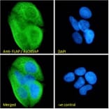
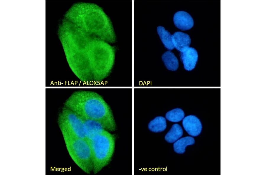
FLAP expression in MCF7 cells analyzed by immunofluorescence. Cells were permeabilized with 0.15% Triton. Staining was performed with Anti-FLAP Antibody (A84031) at 10µg/ml for 1 hour and Alexa Fluor 488 secondary antibody at 2µg/ml. Strong ER staining shown and nuclei were stained with DAPI (blue). Negative control: Goat IgG Isotype Control at 10µg/ml followed by Alexa Fluor 488 secondary antibody at 2µg/ml.
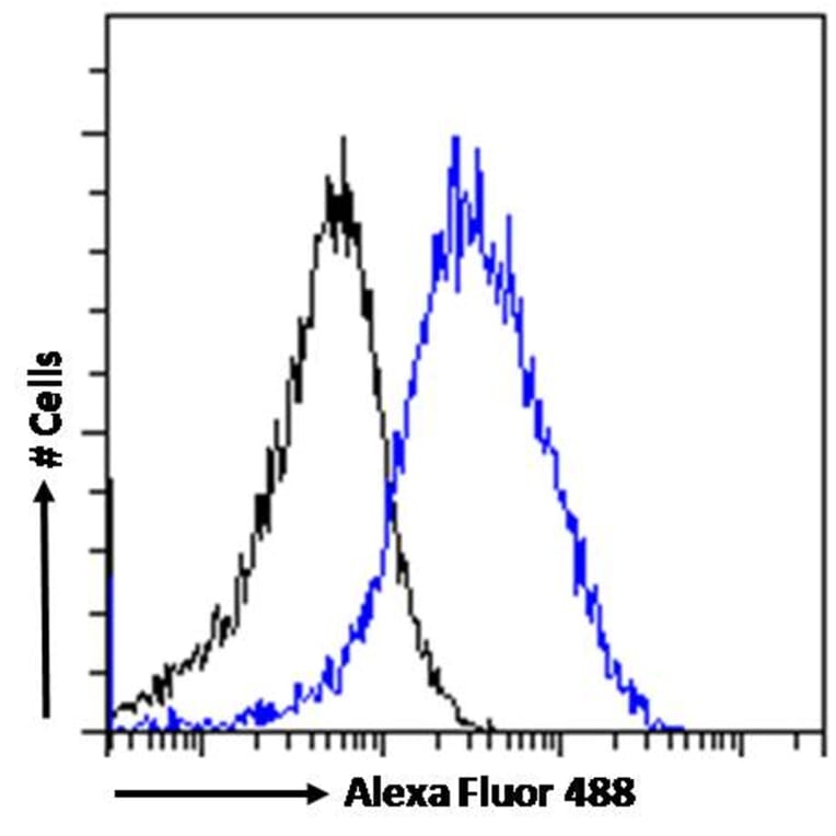
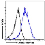
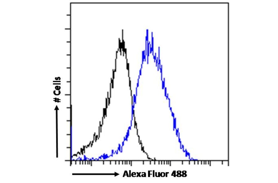
FLAP expression in MCF7 cells (blue line) analyzed by flow cytometry. Cells were fixed in PFA and permeabilized with 0.5% Triton. Staining was performed with Anti-FLAP Antibody (A84031) at 10µg/ml for 1 hour and Alexa Fluor 488 secondary antibody at 1µg/ml. Negative Control: Goat IgG Isotype Control (black line) followed by Alexa Fluor 488 secondary antibody.
Publishing research using Anti-FLAP Antibody (A84031)? Please let us know so that we can list the citation on this page.
Alternative products to Anti-FLAP Antibody (A84031)