Synthetic peptide corresponding to the C terminal region of human GADD34.
Sequence
C-AAALDLSGRRG
Host
Goat
Clonality
Polyclonal
Isotype
IgG
Conjugate
Unconjugated
Purification
Purified from goat serum by ammonium sulphate precipitation followed by antigen affinity chromatography using the immunizing peptide.
Concentration
500 µg/ml
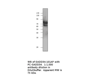
Molecular Weight
75 kDa
Predicted MW
73.4kDa
Product Form
Liquid
Formulation
Supplied in Tris Buffered Saline, pH 7.3, with 0.5% BSA and 0.02% Sodium Azide.
Storage
Shipped at 4°C. Upon delivery aliquot and store at -20°C. Avoid freeze/thaw cycles.
Synonyms
Growth arrest and DNA damage-inducible protein GADD34, Myeloid differentiation primary response protein MyD116 homolog, PPP1R15A, Protein phosphatase 1 regulatory subunit 15A
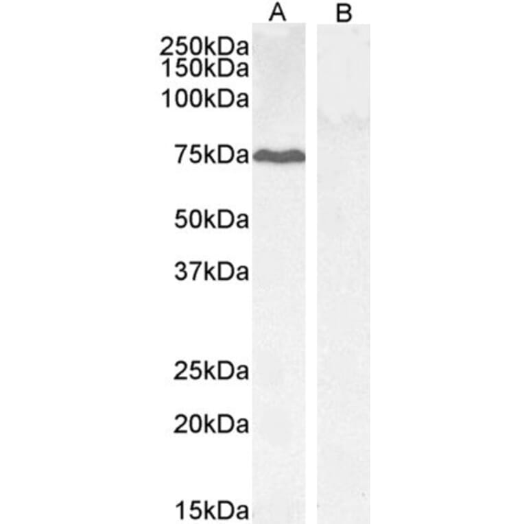
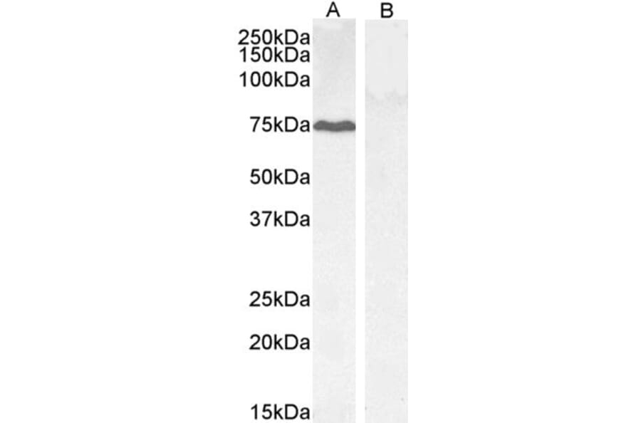
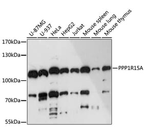
GADD34 expression in HepG2 (A) and negative control KLY (B) lysate analyzed by western blot. Cells were lysed in RIPA buffer and 35µg protein was run per lane. Primary antibody incubation was performed with Anti-GADD34 Antibody (A83758) at 0.3µg/ml and detected by chemiluminescence.
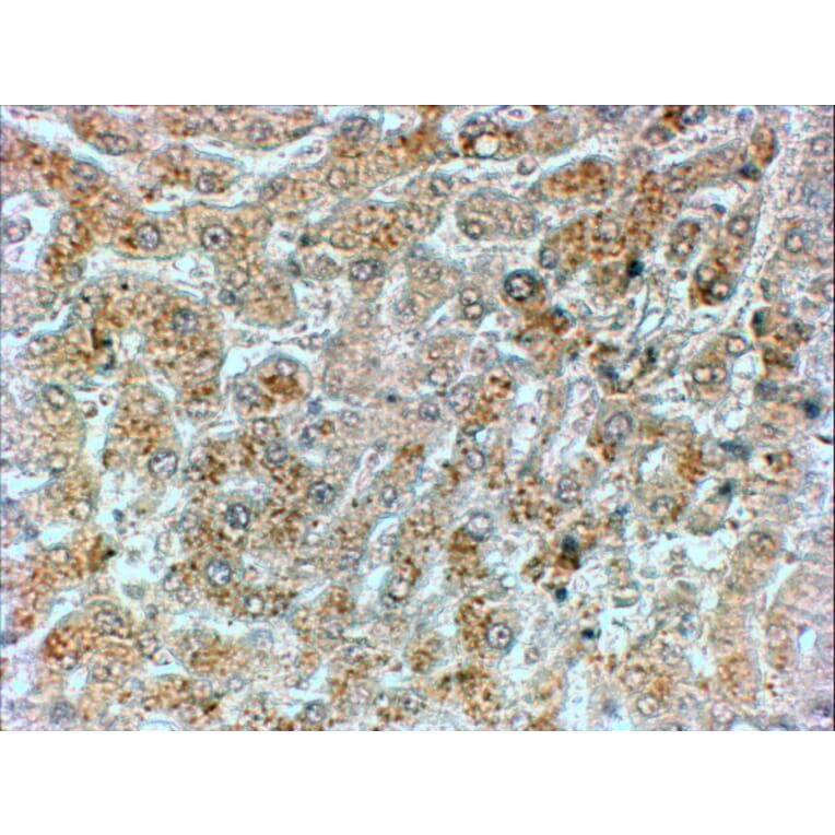
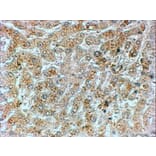
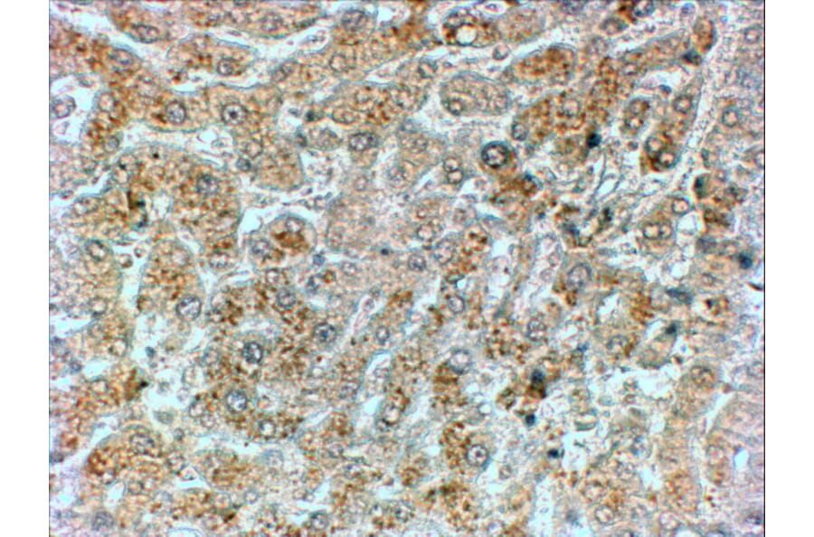
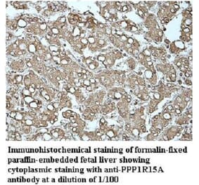
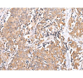
GADD34 expression in Human Liver analyzed by immunohistochemistry. Tissue was paraffin-embedded, and antigen retrieval was achieved by steaming in citrate buffer, pH 6. Staining was performed with Anti-GADD34 Antibody (A83758) at 2µg/ml and revealed with horseradish peroxidase (HRP).
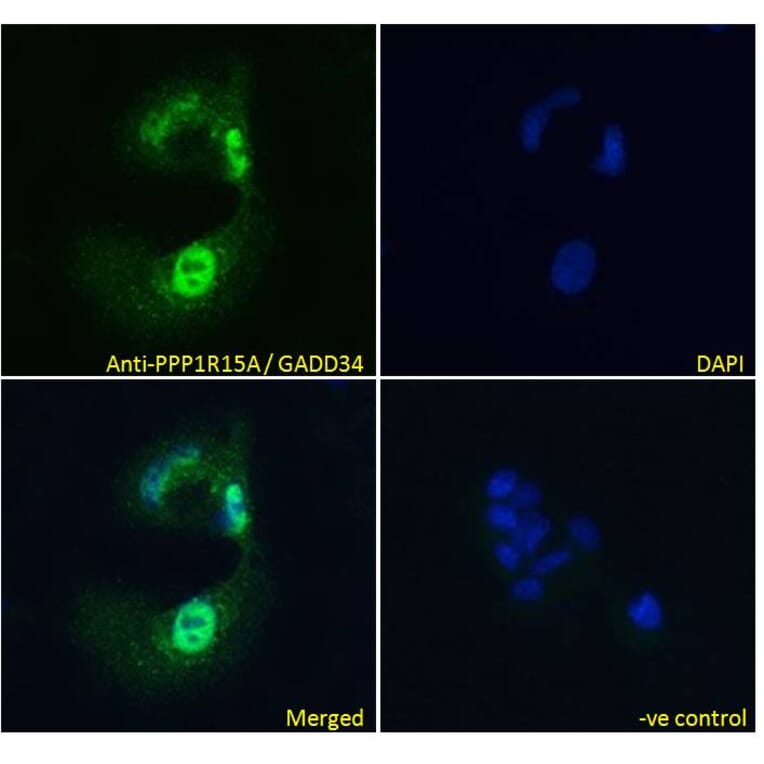
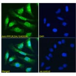
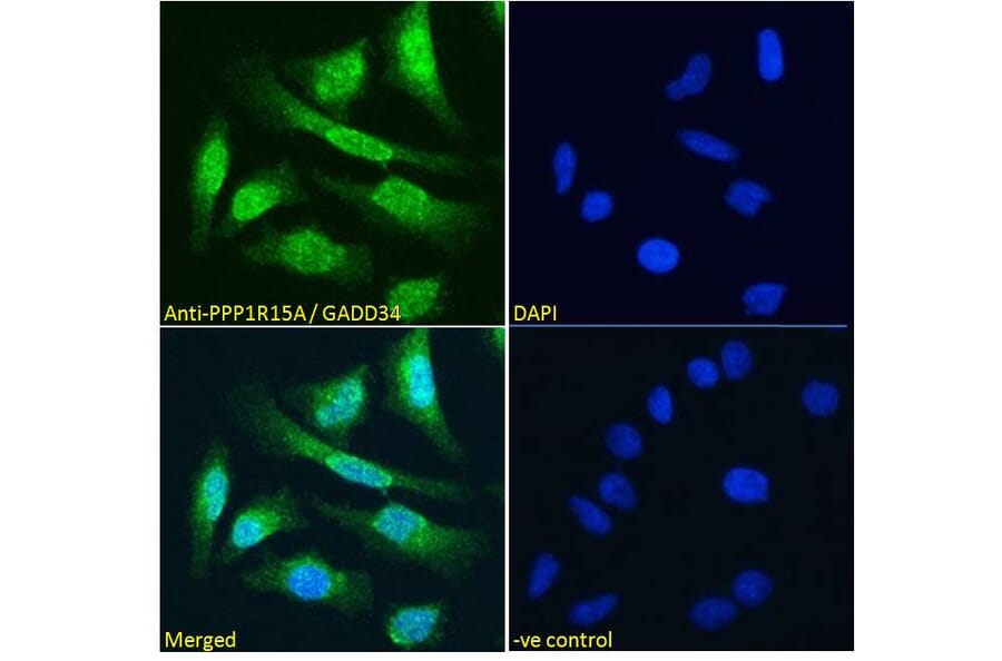
GADD34 expression in HeLa cells analyzed by immunofluorescence. Cells were permeabilized with 0.15% Triton. Staining was performed with Anti-GADD34 Antibody (A83758) at 10µg/ml for 1 hour and Alexa Fluor 488 secondary antibody at 4µg/ml. Some cytoplasmic and strong nuclear staining shown and nuclei were stained with DAPI (blue). Negative control: Goat IgG Isotype Control at 10µg/ml followed by Alexa Fluor 488 secondary antibody at 4µg/ml.
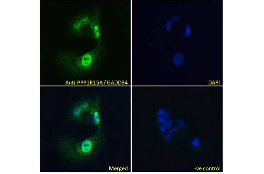
GADD34 expression in HepG2 cells analyzed by immunofluorescence. Cells were permeabilized with 0.15% Triton. Staining was performed with Anti-GADD34 Antibody (A83758) at 10µg/ml for 1 hour and Alexa Fluor 488 secondary antibody at 4µg/ml. Some cytoplasmic and strong nuclear staining shown and nuclei were stained with DAPI (blue). Negative control: Goat IgG Isotype Control at 10µg/ml followed by Alexa Fluor 488 secondary antibody at 4µg/ml.
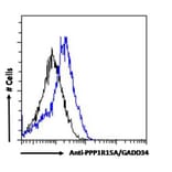
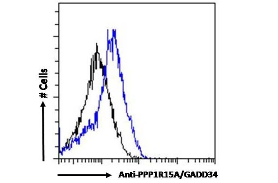
GADD34 expression in HepG2 cells (blue line) analyzed by flow cytometry. Cells were fixed in PFA and permeabilized with 0.5% Triton. Staining was performed with Anti-GADD34 Antibody (A83758) at 10µg/ml for 1 hour and Alexa Fluor 488 secondary antibody at 0.4µg/ml. Negative Control: Goat IgG Isotype Control (black line) followed by Alexa Fluor 488 secondary antibody.